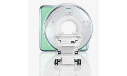
In the near future, diagnosis of cancer and life-saving treatment will mean a two-for-one visit to a physician’s office. At least, that’s the future researchers at Rice University and Baylor College of Medicine (BCM) envision.
Led by Naomi Halas of Rice University and BCM’s Amit Joshi, a recent study has shown that using specially tagged nanoparticles (which were developed by Halas) and an MRI scan clinicians could knock out cancer cells in particular organs at the same time they’re discovered. The research is among the first in a new field called “theranostics” that develops techniques for the diagnosis and treatment of diseases in a single procedure.
MRI is particularly useful as a non-invasive way to track the biodistribution of the nanoparticles as the team moves to testing the technique on animals and, eventually, humans with the ultimate aim of receiving Food and Drug Administration (FDA) approval for the procedure. The current research has used cell cultures.
Clinicians will use an MRI scan to pinpoint diseased tissue, inject the nanoparticles and treat the site with heat, destroying the cancer cells. The nanoparticles include a layer of iron oxide that make them visible to an MRI detector allowing clinicians to observe the particles’ progress through the body in real time.
The researchers see this method as particularly efficacious in treating early-stage cancers. The results of the initial research are available online in the Journal of Advanced Functional Materials.
(Source: Press Release)


