 If you can’t find a decent reason or excuse to attend a major medical imaging meeting in Hawaii, then your priorities are in serious need of adjustment. The International Society of Magnetic Resonance in Medicine (ISMRM) recently held its 10th annual meeting in Honolulu, Hawaii, from May 18 to 23, 2002. This meeting of imaging specialists focused exclusively on the science and medical applications of advanced MRI for a solid six day period, with only an occasional beach break. At least, that’s my official version of the meeting.
So, with my sunscreen, exhibit guide and abstract book in hand, I set out to discover the coming attractions that mainstream radiology departments can expect to hear more about during the run-up to budget season and RSNA this year and next.
3T Systems
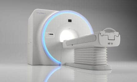
Citius, Altius, Fortius — the Olympic motto of faster, higher, stronger — should be the new standard as high-field imaging has arrived, and you should begin preparing to find a home for your tired, old 1.5T system. 3T is now officially ready to roll, and no respectable prestigious radiology department can afford to do without one if they strive to remain part of the 21st century. 3T systems deliver higher quality images, acquired at faster speeds, with particular application to brain and cardiovascular imaging.
Beginning with the opening session, almost all of the main presentations at ISMRM focused on high-field imaging. The technological challenges associated with this increased field strength primarily revolve around control of the static and switched fields and RF energy levels that must be controlled for patient safety. Solutions to these challenges are well underway, which will then leave users with significant benefits, primarily improved spatial resolution (from higher SNR), faster scan times and new imaging protocols (including spectroscopy applications) for advanced studies.
Since this will be the first generation of 3T for mainstream clinical use, expect that the rate of change (need for upgrades) will be fast for this imaging platform, as coils, pulse sequences, image processing SW and gradients are each modified and improved to further harness the raw power of these systems. You may want to plan a healthy budget for upgrades during the first few years if you decide to jump on this bandwagon anytime before 2004.
You should expect to see an initial wave of orders as the early adopters — with plenty of budget and research to accomplish — put these systems through their paces of the routine and rare cases. There is no need for a financial justification for clinical use of these systems — yet. There is a compelling confidence that the clinical world of brain disease will receive a tremendous boost from the use of these systems to better characterize disorders that require both metabolic and anatomic evaluations.
Squiggly Lines
Have you ever looked at MR spectroscopy (MRS) studies? Well, you’re not alone if you haven’t. In essence, with MRS you are looking at bio-chemical data, not the usual sharp high contrast anatomical image that is the normal result from an MRI system.
If you work in a radiology department, you are definitely ahead of most groups if you already make regular use of MRS to diagnose neurological diseases and disorders. The clinical utility is well proven, the reimbursement is in place, the benefits for patient care are solid, and the techniques are push-button. Now we just need more education of physicians (and especially radiologists) in order to broaden the use of these procedures. Take some time to improve your knowledge of this area, as it is becoming an even more important part of the MRI portfolio for state-of-the-art work.
Even Higher Fields
As 3T systems begin their move from the clinical research frontier into mainstream clinical use, there is (obviously) something to take its place. Make room for the new 7T and 9T MRI systems, which are now beginning to take hold in the basic research world. MRI is now making its way into drug research and trials, even pre-clinical work with animal studies.
Memo to Grads – Physics!
Given the paltry graduation rate of advanced degrees for physics majors in the U.S., we need to generate some more interest in this subject area as the expanding world of MRI has a tremendous need for more people ready to take this science to the next level. So pull aside some of the top graduates from your neighborhood high schools, and encourage them to consider this field. At first glance, physics looks like textbook-bound science. But you might make a difference for some of these future MRI scientists by explaining how the world of advanced medical diagnosis depends on the continuing work of MRI research, which is almost completely led by the science of physics and medicine.
Doug Orr, president of J&M Group (Ridgefield, Conn.), consults with medical device companies in strategy and business development for emerging growth markets, notably radiology and cardiology. Comments and suggestions can be sent to [email protected].


