Yes, it is still impractical and costly, but when available, for the right patients, physicians continue to see value in PET.
 |
Positron emission tomography (PET) has been around for decades and is finding a clinical foothold with oncology and research. But PET has always been the ugly stepsister to single photon emission computed tomography (SPECT) when it comes to cardiology applications. Physicians and hospital administrators see PET as expensive, cumbersome, and completely impractical for the emergency department.
At the same time, cardiologists fortunate enough to have a nearby cyclotron or a tabletop rubidium-82 generator say that—for some patients—there is nothing like PET … for now.
The Upsides of Cardiac PET
Furqan Tejani, MD, cardiologist, director of advanced cardiovascular imaging at Long Island College Hospital, Brooklyn, NY, is one of PET’s true believers. Despite the major PET manufacturers reducing or eliminating their cardiac PET packages and PET/CTs sales being flat, Tejani does not see cardiac PET dying a death of attrition.
“The strength of the radioisotopes used in PET is many orders of magnitude over and above what you use in SPECT or in standard nuclear cardiology,” Tejani said. “So it gives you images that are much more efficient, much better, and images that are much more representative of the true physiological review of the cardiac muscle fibers, versus SPECT, which generally gives you an approximate to a fairly gross idea of what’s going on.”
John A. Rumberger, MD, PhD, director of cardiovascular imaging at the Princeton Longevity Center in New Jersey, said that PET’s clarity in perfusion mapping is a huge advantage over SPECT.
“It’s like looking at something in a dim light versus looking at something in a bright light,” Rumberger said. “All of a sudden, instead of saying, ‘I’m kind of sure I see something,’ it’s like someone turning on the light bulb in the room, and now you say, ‘Oh, OK, there it is.’ “
Marcelo F. DiCarli, MD, chief of nuclear medicine and molecular imaging, director of cardiovascular imaging, Brigham and Women’s Hospital, Boston, added: “PET has quantitative capabilities for measuring and estimating coronary vascular reserve in the coronary tree, which enables better detection of patients with balanced ischemia and multivessel disease.”
Along with evaluating cardiac viability, cardiologists also appreciate PET studies for its help guiding surgical treatment decisions. Tejani said, “If you’re looking at areas of the myocardium that are potentially revascularizable, or if you’re looking at somebody who is being explored for cardiac transplantation and you want to make sure that there are no areas that are revascularizable, then this is where PET really becomes useful.”
DiCarli noted that the latest and greatest PET/CTs enable physicians to collect accurate anatomic information in those with coronary disease in the form of a calcium score or a CT coronary angiogram. Unlike stand-alone PET, a PET/CT can provide reliable attenuation correction, which can lead to improvements in specificity, avoiding false positive studies.
“Utilizing PET/CT provides an opportunity for a very comprehensive evaluation of both the anatomic extent of disease and its physiologic significance,” DiCarli said. “This should allow us to better characterize the process and the severity of coronary artery disease and also to assess patient risk by defining both the amount of ischemia as well the extent of anatomic disease.”
The Downsides of Cardiac PET
PET is often described as one of the more “elegant” imaging techniques, but all of that elegance and sophistication comes at a cost.
Cyclotron radiopharmaceuticals for cardiology may provide the most refined images with F-18 fluorodeoxyglucose and nitrogen-13 ammonia studies. But cyclotrons are not installed in every city.
The availability of PET cameras, as well as radiopharmaceuticals, is a limitation compared to SPECT,” DiCarli said. “So, in order for cardiac PET to really be able to succeed, we need to have a model of a unit dose distribution of radiopharmaceuticals similar to what FDG has done for the oncology PET market. But we’re not there yet.”
Consequently, most cardiology PET centers rely on tabletop generators that utilize strontium to generate rubidium-82 for cardiac PET studies. While rubidium generators have certainly made PET more convenient with less dependence on a cyclotron, the PET protocol is still complicated.
“It’s not that a good study can’t be done [with rubidium], but it requires very significant diligence in doing it right,” Rumberger said. “If you do it right, it’s useful. But if you inject too late and don’t do it right—maybe you’ve waited too long for the strontium to make the rubidium—then it’s lost its energy, and then the test isn’t very good.”
Another challenge is that rubidium is not always the ideal radiopharmaceutical for every patient, Rumberger said. “Rubidium has a limitation in terms of the increases in blood flow that can be seen. In the average normal person, if we were to give you a drug that would dilate your heart arteries and increase the blood flow to your heart, it would go up about 4 to 6 times normal. With rubidium, anything above about 2 and a half to 3 times normal would look the same. So it has an upper limit of its potential ability.”
Then there is the extra expense. PET/CTs allow for some flexibility in that one can utilize the CT function without PET, but in the age of tighter reimbursements and rapid technology obsolescence, CFOs must consider the extra expense for PET functionality and the number of studies that might actually be performed—and covered by insurers. Some insurers still consider cardiology PET exams experimental relative to SPECT, while others will approve and cover it on a case-by-case basis.
On top of all that, PET/CT cardiology studies generally have a higher radiation exposure compared to a SPECT stress test or a single coronary CTA. Consequently, even if costs could be lowered for the scanner, PET and PET/CT will never become a routine screening study, but remain a tool for higher-risk patients. That limits patient volume, which in turn limits a financial return on investment for a typical cardiology imaging suite.
To PET or Not to PET
Despite all of PET’s limitations, when available and covered by insurance, some physicians prefer PET for particular patients because of its superior image quality compared to SPECT.
“At the moment, if both SPECT and PET are available at an institution, I think that PET offers the possibility of improved diagnostic accuracy and efficiency compared to SPECT for patients who need a pharmacological stress,” DiCarli said. “Usually, patients who need a pharmacological stress are patients who tend to be more obese or are unable to exercise. They tend to be sicker patients, in whom, unequivocally, you get better image quality for PET compared to SPECT.”
However, for those patients who are able to get on a treadmill, SPECT is preferable. “Robidium only allows us to do pharmacological stress testing because of its very short half-life. Therefore, we cannot couple PET imaging with exercise, which is a disadvantage, compared to SPECT,” DiCarli said.
Aside from PET being more desirable because of its improved sensitivity and specificity, there is also the time savings. PET cardiology studies take around 30 minutes compared to approximately 3 hours for a full SPECT study.
PET and PET/CT are not radiation free, and the risks must be balanced versus the benefits for a particular patient. Due to their expense, time, and cumbersome nature, PET modalities are certainly not a cardiac screening exam. But for higher-risk patients heading into surgery, it can certainly be worth the risk.
“If you have a high-risk patient with a high-risk procedure, that’s perfectly acceptable, because at the end of the test, you’re getting a very high degree of information,” Tejani said.
While concerned with radiation, DiCarli said that the latest PET technology is comparable to SPECT. “In general, radiation dose symmetry from PET imaging is somewhat lower than SPECT, though it really depends on the radiopharmaceutical that you use. If you’re using rubidium-82, it’s quite comparable to a single-day SPECT study. If you’re using another flow agent like nitrogen-13 ammonia, the PET dose is about a third of the dose that you would get from a SPECT study. So it really depends. At worst, it’s about equal to SPECT. At best, it can be a third of what you can get from SPECT imaging.”
The Future of PET and Cardiology
Because of PET’s cost and impracticalities, and less cumbersome alternatives, many see PET fading away as a standalone modality for cardiology. For the most part, the major PET players have seen the writing on the wall and have discontinued their stand-alone PET offerings in favor of PET/CT.
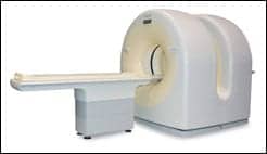 |
| Philips’ Gemini PET/CT features time of flight technology. |
But even true cardiac PET believers such as Tejani expect that new products such as Tustin, Calif-based Toshiba’s new 320 multirow CT may one day fill that cardiac perfusion role—but not yet.
“CT imaging, in due course of time, will become the de facto standard for perfusion imaging,” Tejani said. “But at this point, SPECT isn’t mature enough and robust enough of a technology and PET has the capability of doing perfusion, not only elegantly, but also doing it scientifically accurately. So, I believe PET will stick around for a while until CT matures and becomes the standard for perfusion imaging.”
DiCarli agrees that CT is a long way from becoming the go-to for cardiac perfusion imaging. “Other than the animal data, there’s not even a shred of data to suggest that this is clinically viable,” he said. “So if and when—and we’re part of that [Toshiba] clinical trial—if and when we can measure tissue perfusion with CT, then we need to find out what the role of CT may be there. But I think that discussion is hypothetical at this point.”
Cardiac MRI techniques are also maturing. But Tejani does not view these as being a replacement for PET, either. “The only advantage to MR is that there is no radiation involved and that MR could be done at any odd time. So PET does require a significant amount of logistics around it, but by the same token, MR is just as cumbersome.”
Asked about prospects of improving the PET modality so that it becomes more practical and more widely available, Tejani does not see that in PET’s future.
“PET is a very elegant imaging modality, which involves a significant amount of physical principles that are either being altered or followed. So, it’s very difficult to say that there will be a revolution in PET imaging and that all of a sudden there will be a PET that is completely different, or an isotope that’s not going to be the way it is. It’s not going to happen.”
Tor Valenza is staff writer for Medical Imaging. For more information, contact .