Father-and-Son Radiologists Look at Changes Over 40 Years and Those to Come
Product Showcase
Running the Numbers
Father-and-Son Radiologists Look at Changes Over 40 Years and Those to Come
Since 2002, Philadelphia magazine’s Top Docs feature has recognized two Wallace Miller, MDs—father-and-son radiology duo Wallace Miller, Jr, and Wallace Miller, Sr, who practice side-by-side at the Hospital of the University of Pennsylvania (Philadelphia). Miller, Sr, has been practicing since 1962; in 1991, his son joined the team.
“I’m my dad’s prot?g?,” Miller, Jr, said. “We work very similarly.” Miller, Sr, agreed, saying, “One thing I’ve always felt about reading films is that you should stick out your neck and make a diagnosis, try to be positive—and not always mention extraneous things that probably are not important. And I think Wally does that as well as anybody else.” He added, laughing, “Outside of me.”
 |
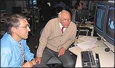
| The father-and-son duo of Wallace Miller, Jr, (left) and Wallace Miller, Sr, (right) work side-by-side as chest radiologists at the Hospital of the University of Pennsylvania. According to Miller, Jr, “Occasionally, my dad and I will disagree slightly on the interpretation of an exam. Everyone thinks that?s hilarious, a family spat. But in reality, that?s the way life is?people who come from similar backgrounds can ultimately have different opinions.” |
When Miller, Sr, started as a resident, films were still developed by hand, and turnaround took as long as a week—film dried overnight, was read the next day, and the radiologists’ dictation was typed for eventual transmittal to the referring physician. “They found out about their patients by coming down and talking to us,” he said. “Basically, a report didn’t get to them for days. Now, we send the report back to the referring physician before the patient gets back to the doctor’s office.
“There is one disadvantage to that,” Miller, Sr, continued. “Whenever the referring doctor didn’t get the reports, he came down and talked to us. Now, he gets a report and can actually look at the films on a monitor in his office. I think we lose in not having that interchange, and I think the patient loses a little bit, too.”
Miller, Jr, practices chest radiology in the same department as his father, reading chest x-rays, chest CTs, and the occasional PET scan. Both agree that chest CT faces a risk of being overused when frequently, chest x-ray is sufficient. “People don’t work out things on the chest x-ray the same way they used to,” Miller, Jr, said. “My dad is a master at it.” Miller, Sr, added, “Unfortunately, a lot of people say, ?There’s something abnormal here; let’s get a CT.’ I think a lot of unnecessary CTs are done.”
Asked whether the practice of chest radiography was eroding, Miller, Sr, said, “You can get an awful lot of information out of a chest x-ray.” But Miller, Jr, while acknowledging that chest x-ray could still play a significant role in diagnosis, disagreed: “It’s clear in my practice that that’s already started to begin.? The chest radiograph was the core of the evaluation of a pulmonary disease, and it no longer stands like that.”
The two physicians also have different visions of the future of radiology. “When I was a resident, I had no idea that things were going to develop as they did,” Miller, Sr, said. “We thought we were pretty good at reading chest x-rays, but we had no idea that one day, we’d be looking at ultrasounds, CTs, MRs. I have no idea what the future’s going to be like, but I predict that some new modality will come along and will probably revolutionize the way we look at things, even as CT and MR have revolutionized things during my lifetime.”
Miller, Jr, offered a slightly different perspective. “I think that anatomic imaging has become a mature practice. There will be incremental changes in the future, but there aren’t going to be any major changes, I don’t think, except that we’re going to be able to see things smaller and smaller. So, what I think most people are predicting is that the next big thing in imaging is going to be functional imaging—looking at the body from an imaging perspective, but based on the physiology and the microbiology of the body. We’ll see if we succeed. But I think that’s where we would like to go.”
—C. Vasko
Product Showcase
Digital X-ray Image Detector Produces 30 Images Per Second
Varian Medical Systems Inc (Palo Alto, Calif) recently announced the introduction of its new PaxScan 1313 image detector. Optimized for use in dental and orthopedic imaging as well as with semiconductor inspection systems, the PaxScan 1313 features a 13- x 13-cm imaging area and can produce up to 30 images per second.
 |
| The PaxScan 1313 is designed to be more affordable and offer better image quality than image-intensifier tubes. |
“Image-intensifier tubes offer only 8-bit depth, they degrade over time, and they must be replaced frequently in some cases, raising their cost,” said Gary Okamoto, imaging products marketing manager at Varian Medical. “Image quality is better with the PaxScan detector. It generates images of up to 16,000 shades of gray, thanks to a 14-bit depth analog-to-digital converter.”
The PaxScan 1313 also boasts a 127-?m pixel pitch and a cesium iodide scintillator optimized for scanning at 80 to 90 kV to minimize dosage; each panel comes with ViVA software, a CameraLink connector for digital video output, a universal compact power supply, an optional image-capture board, and cables to cover multiple applications.
Running the Numbers
30% more revenue in 2005 is part of what positioned Trixell (Totowa, NJ) for the 2006 Frost & Sullivan Award for Global Excellence in DR detector technology. By sales volume, Trixell?a joint venture created in 1997 by Thales (Totowa, NJ), Philips Medical Systems (Andover, Mass), and Siemens Medical Solutions (Malvern, Pa)?is the leading manufacturer of flat-panel digital detectors. The company sold nearly 3,000 detectors in 2005 alone, accounting for half of the world?s flat-panel detector sales. For more information, visit www.frost.com.