|
Caring for pediatric patients can be a complex process. Differences in the physiological and anatomical characteristics of children require meticulous attention to detail and patient management, from presentation to treatment. Pediatric diagnostic imaging differs from adult diagnostic imaging in a number of ways, including altered radiation dosing, different immobilization needs, and changes in certain technical factors affecting the production of images of very small, developing physical structures.
Those providing pediatric radiology services frequently deal with children who may be in pain, irritable, or lethargic from injury or illness. Therefore, the range of challenging behavior that these children may display must be managed if the department or imaging center is to produce high-quality images from which accurate diagnoses can be obtained. Radiology personnel must provide, as much as possible, a nonthreatening environment, along with sufficient reassurance to calm the patient. Frequently, parents must be present to hold or comfort their children. This situation can add to the complexity of the radiologic technologist’s work.
Depending upon the study to be performed, parents may need to be provided with dietary instructions for their child. If the mother or other female caregiver is pregnant, it should be communicated to the family that the father, or another nonpregnant relative, should accompany the child to the examination. All procedures should be explained to the parent or parents and to the child, if he or she is old enough, in language that the child can understand.
INJURY AND SAFETY
Injury is the leading cause of death among children who are 1 or more years old 1 ; likewise, many radiology departments perform a large number of injury-related studies of children. Between 30% and 45% of children who have experienced trauma have multiple injuries, including at least one skeletal fracture. 1 Given this level of fracture prevalence, evaluation of each extremity is necessary. Radiographic evaluation of the cervical spine, chest, and pelvis has become the baseline norm employed in assessing injured children. Because of the normal anatomical variations of children, as compared with adults, only experienced pediatric radiographers should perform these studies. Additional studies may be needed, depending upon the nature and mechanism of the injury.
Children’s development can be affected by excessive exposure to radiation. The amount of radiation delivered to children during radiographic examinations, however, is less than that required for adult imaging.
It may be necessary to restrain or sedate children who are undergoing imaging examinations, both to ensure that the areas under examination are irradiated properly and to avoid irradiating areas other than those necessary for the diagnostic study that has been ordered.
EQUIPMENT
Digital radiography (DR) allows pediatric imaging departments and centers to avoid the need for retakes because images can be manipulated at the workstation. The ability of the radiologic technologist to optimize the image on a monitor not only improves throughput and departmental productivity, but also ensures that ill and injured children will not have to remain in the examination area for any appreciable length of time. In addition, no retakes means that a much lower amount of radiation is delivered to the patient. This is particularly critical for the safety of pediatric patients.
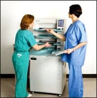
 The design of Swissray ddR is particularly child-friendly. The design of Swissray ddR is particularly child-friendly. |
Equipment for pediatric DR comes in a variety of configurations. The Swissray direct digital radiography (ddR) system allows for maximum flexibility in positioning, since an examination table is not needed and the C-arm design makes the unit less obtrusive than other configurations. When it is necessary to do so, parents are able to hold their children with greater ease.
The Swissray ddR system also allows images to be manipulated by physicians at remote workstations. The ability to isolate and enlarge a particular portion of an image has an added benefit in interpreting studies of children because it makes it possible to examine smaller and developing anatomical features in detail to ensure that nothing is missed.
STRATEGIC PLANNING
Many hospitals, including pediatric institutions, have entered the world of digital radiology, often with the object of creating a highly accessible system for clinicians while eliminating film and its attendant costs. In addition, they are typically able to reallocate the space used for traditional radiography equipment for other revenue-generating purposes.
According to Linda Wright, radiology director at Denver Children’s Hospital, her hospital entered the world of ddR in 2001 with the implementation of a picture archiving and communications system (PACS). A natural next step was to move from film to filmless imaging to improve productivity and reduce costs over the long term. The hospital introduced two Swissray ddR units to coordinate with the implementation of the PACS, and the facility is now 98% filmless.
CONCLUSION
Pediatric imaging must take into consideration the special needs of its developing patients. Equipment-purchasing decisions must be made with safety and consumer friendliness as top priorities; ddR has the potential to meet these needs in a way that also optimizes the financial return to the health care facility.
A rational approach to DR makes sense in terms of reducing overall costs and increasing ease of implementation through the coordination of software and hardware purchasing and installation. Clearly, DR technology should be considered as part of the overall technology strategy of any health care enterprise.
Marilyn Ferdinand is a contributing writer for Decisions in Axis Imaging News.
References:
- Nguyen TD, Raju R, Lee S. Considerations in pediatric trauma. Available at: www.emedicine.com. Accessed October 22, 2004.