Prostate cancer is receiving more attention than ever — driven by well-informed patients and by the cancer’s prevalence. The second leading cause of cancer death in men, prostate cancer will strike one man in six at some point in his life, according to the American Cancer Society (ACS).
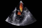
The prostate-specific antigen (PSA) test gives doctors an accurate early warning sign that prostate cancer is present. But once cancer or other prostate disease is suspected, imaging the organ and its surrounding tissue to determine the type, location, and extent of disease remains difficult. Most physicians agree that transrectal ultrasound is the first step in looking for cancer.
After that, physicians differ in their imaging approach.
For many patients, an ultrasound biopsy confirming cancer is the only imaging test they receive before treatment. Others also receive tests to look for metastasis. Physicians have their choice of computed tomography (CT), magnetic resonance imaging (MRI), positron emission tomography (PET) or single-photon emission computed tomography (SPECT). Although physicians disagree about the best approach, most agree that none is as good as they would like.
Some physicians say improving prostate imaging lies in looking beyond the anatomy to evaluating organ function. And we are nearly there, they say.
Imaging challenges
“Imaging the prostate is a difficult undertaking,” says Claus Roehrborn, M.D., associate professor of urology at the University of Texas Southwestern Medical Center (Dallas). Hidden under the bladder and pubic bone, the prostate’s location is one complicating factor. In addition, because the prostate cannot be filled with contrast, it is impossible to image by standard X-ray, Roehrborn says.
Please refer to the September 2001 issue for the complete story. For information on article reprints, contact Martin St. Denis