Clinical benefits, added diagnostic assurance, and patient requests for the technology are keeping CAD for mammography in demand.
The primary benefit of computer-aided detection (CAD) for breast imaging, according to Matthew Gromet, JD, MD, section chief of breast imaging at Charlotte Radiology (Charlotte, NC), is its ability to assist the radiologist in avoiding oversight of a significant lesion.

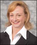
Matthew Gromet, JD, MD, Section Chief of Breast Imaging, Charlotte Radiology
He mentions, for example, a subtle abnormality that a radiologist may overlook. In these instances, he says, CAD may highlight that area and cause the radiologist to take another look to see if it is significant. “The goal of CAD for breast imaging,” said Gromet, “is to increase sensitivity in finding early breast cancer.”
The screening environment is where CAD is the most useful, says Gromet, who uses R2 ImageChecker Digital CAD from Hologic (Bedford, Mass). “You’re reading screens with one decision to make: ‘normal’ or ‘recall for further assessment,'” he said.
Clinical Benefits
Having used CAD for about 5 years, Randy Hicks, MD, MBA, CEO at Regional Medical Imaging (Flint, Mich), agrees with Gromet that it is an invaluable tool in breast imaging. He notes that his practice—one of the American College of Radiology’s Centers of Excellence in Breast Imaging—does a lot of mammogram screenings.
“[As a radiologist,] you’re doing the same thing over and over again, so a second eye—a second-look opinion—is helpful. [Using CAD is] almost like getting a dual read,” said Hicks, who uses R2 ImageChecker Digital CAD from Hologic, CADStream from Merge Healthcare (Chicago), and SecondLook Digital from iCAD (Nashua, NH).
“Probably the biggest utility with CAD is in finding microcalcifications,” said Hicks, who notes that CAD is much more sensitive than the human eye. However, when it comes to findings of masses and architectural distortion, he finds CAD less helpful. “[CAD] doesn’t look back at previous years,” he said. “It looks at a single shot in time.”
When it comes to finding cancer in general, CAD is “invaluable,” he said. “CAD has saved lives and has found cancers we would have missed,” said Hicks, who notes that, for a minimal cost, CAD adds value, and he suspects that, eventually, CAD will be integrated into all mammographic systems.
Marcela Böhm-Vélez, MD, FACR, a radiologist at Weinstein Imaging Associates (Pittsburgh), says that the number of markings might distract residents who are just learning how to interpret mammograms, but she believes CAD also can be helpful to residents, since it can “serve as a great training device.” Fellows also benefit from working with CAD, she believes, since the software can give them extra confidence as they navigate through mammograms. General radiologists who interpret mammograms—in addition to other types of diagnostic images—also can stand to benefit from the technology, according to Böhm-Vélez.

Marcela B?hm-V?lez, MD, FACR, Radiologist, Weinstein Imaging Associates
She agrees with Hicks that CAD is much better for calcifications, while admitting that the technology is not as effective at accurately detecting masses. “My eyes get tired. CAD does target me to calcifications that, hopefully, I wouldn’t miss.
“Calcifications are a sign of very early cancer—and we want to detect cancers early because the survival rate for patients [at that stage] is between 96% and 97%,” she said.
“I’m pretty good at finding microcalcifications, but [CAD] would beat me every time,” concurred Robert Schmidt, MD, professor of radiology (retired) at the University of Chicago, who believes that the technology helps him find areas of the breast where he should focus his attention as he looks for abnormalities. “CAD generally finds 98% of calcifications, many more than any radiologist would find on their own.”
Reimbursement’s Role in Adoption
While providing the best care to patients should always be of first concern, Schmidt believes that reimbursement is often key to physicians’ adoption of new technologies. This is “difficult in a new era of medicine where doctors are controlled by the reimbursement process,” he said.
“The medicine that will get done is the medicine that is paid—not the medicine that’s best for you. I don’t think we can get away from this, though there is a subtle underlying pressure to do things more economically,” said Schmidt, who uses R2 ImageChecker Digital CAD from Hologic.
But in the case of CAD, reimbursement is not the key driver of adoption. Schmidt concluded that “the reimbursement for CAD—something like $18—could disappear and radiologists would still use it. Namely, because CAD really improves radiologists’ efficiency—otherwise, they wouldn’t use it.”
Driven by Patient Demand?
Böhm-Vélez, who uses SecondLook Digital from iCAD and Volpara for Volumetric Breast Density from Matakina Technology (Wellington, New Zealand), says that she was driven to get CAD because of her patients.
“When CAD came out,” she said, “the message was out there that the computer could detect additional cancers. Patients continued to ask me, ‘Do you have CAD?'”
Women asked that question because they wanted to know about their breast density, says B?hm-V?lez. She notes that approximately 50% of women have dense breasts, and, for these patients, additional imaging such as ultrasound could be helpful.

Robert Schmidt, MD, Professor of Radiology (retired), University of Chicago
The breast density issue is very real, says Hicks. “CAD is very much like humans. It has a very difficult time discerning masses, like we do. When you have all white, it’s hard to see a subtle change in the white pattern. But CAD is brilliant at finding calcifications—more so than humans [are]. By the same token, I think I would warn that if CAD is looking for calcifications in a dense-breasted woman, there’s decreased value added because of reduced sensitivity,” he said.
“Radiologists need a lot of help when judging the breast density,” said Schmidt. “I don’t want to insert my opinion on breast density when the computer does a better job. Radiologists always overestimate the amount of density in the breast.”
A significant fraction of patients ask about breast density, says Schmidt. Patient requests for CAD and information on their breast density derive, in part, he says, because companies making CAD systems and alternative screening tools educate the public about its importance.
“It’s a rare week that you don’t see a newspaper article that covers breast density,” he said, while noting that he consults mostly with diagnostic—rather than screening—patients. “They’re alerted [to the availability of CAD for breast imaging], and their doctor has to be knowledgeable [about CAD and other technology advances].”
Hicks acknowledges that patients have access to information about new technologies much more so than ever before, but he doesn’t believe that this impacts his practice’s technology purchasing decisions. “Ninety-seven percent [of patients] still do what their doctor says. They trust Dr Randy Hicks. It isn’t about technology. It’s still about relationships,” he said. Over the course of his 26 years as a radiologist, he has come to believe that the relationship doctors have with patients is what matters to women.
Room for Improvement
As far as Schmidt is concerned, “you shouldn’t even refer to CAD as providing ‘computer-aided diagnosis.'” He prefers to refer to CAD as providing “computer-aided detection” for mammography. “The fact is, [CAD] isn’t usurping power from any [radiologist] reading,” he said.
In fact, he likens the use of CAD for breast imaging to the use of GPS while driving. “If the GPS tells you to move left and there’s a brick wall [on your left], you’re not going to turn. So, radiologists are still in control of [their interpretations of mammograms],” he insisted.
While he, too, embraces CAD in his practice, Gromet acknowledges that CAD does present reading challenges due to its high marking rate. “Most marks will not be significant. Radiologists must use their skills to dismiss false marks, while at the same time recognizing potentially significant lesions,” he said. “Doing so will avoid a significant increase in the recall rate of false-positive screening mammograms.”
One of the limitations of CAD, says Gromet, is it doesn’t find all lesions. “A radiologist can’t lessen scrutiny of a mammogram because CAD doesn’t mark something. About 21% of invasive cancers recalled by radiologists in our practice were not marked by CAD,” he said. Although he insists that CAD definitely helps, he said that—even with CAD—radiologists “still have a challenging task to do when interpreting women’s mammograms.”
“When it comes to mammography,” said Hicks, “you’re doing it over years and looking for changes. CAD looks at this year and finds what looks like a distortion—but it’s not. Sometimes, you can spend a lot of time double-checking the CAD system to make sure it’s seeing something real.”
Next Generation of CAD
Schmidt harkens back to a time when doctors relied on only their hands to feel for breast lumps. “With technology [such as CAD], we’re able to isolate lumps that are smaller and smaller as we get better over time,” he said.
But he yearns for more than incremental improvements that he has seen over time with CAD for breast imaging. He says that CAD vendors need to consider how they will tackle the following questions: How can we use CAD with new modalities? What about developments in MRI and ultrasound? How do you integrate all of these new technologies? Also on Schmidt’s wish list would be computer analysis of lesions’ character and malignant potential.
According to Hicks, “CAD could be programmed to give quantitative density scores for each mammogram. This information could be combined with [computer-generated] breast cancer lifetime risk assessment scores, which we perform for all routine mammography patients at Regional Medical Imaging.
“Depending on her risk assessment and density scores, a woman would pursue a particular imaging pathway during the course of her life, based in whole or in part on intelligence gathered from CAD for breast imaging,” he said.
Given the increasing focus on individualized medicine, Hicks predicts that, in coming years, radiologists will be able to use CAD to treat patients as individuals. He feels that the ability to provide such an individualized approach to care could be particularly helpful to women with dense breast tissue.
In the future, Gromet expects to see CAD algorithms that are capable of comparing right and left breasts, as well as to prior films. For example, the presence of multiple similar clusters of calcifications in both breasts—or calcifications that haven’t changed over time—may allow the computer to lessen its “suspicion score” of the finding, according to Gromet.
Aine Cryts is a contributing writer for Axis Imaging News.


