 |
· DIRECTVIEW Upgrades!
· Tech Zoom: Planning to be Paper-Free
· Software Solution: Pushing the Envelope in Preoperative Planning
DIRECTVIEW Upgrades!
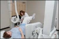 |
| Jennifer Sheftick, RT (RN), prepares to capture a C-spine procedure using Springfield Clinic’s KODAK DIRECTVIEW DR 7500 System. |
Over the course of the past month, Carestream Health Inc, Rochester, NY, has vamped up its KODAK DIRECTVIEW DR 7500 System with new features.
In October, the company announced new software designed to provide more consistent image presentation and orthogonal control of image processing.
“Because it produces more consistent image rendering of varying images, the new EVP Plus software can improve diagnostic confidence,” said Todd Minnigh, worldwide director of marketing for DR. “Better management of noise allows users to optimize their exposure techniques.”
According to Carestream, the software uses a sophisticated algorithm that enables control of brightness, detail contrast, and latitude, among other parameters. Images can be calibrated to radiologists’ preferences using a Look Selection Tool, and the software supports an unlimited number of body parts and projections. Furthermore, a customizable user interface and 45 new electronic image markers improve communication among users. Multiple frequency bands were included to enhance anatomic structures without adding noise.
“EVP Plus software is targeted at improving both productivity and patient care since health care providers need—and have come to expect—continuous improvements in both areas,” Minnigh said. “The primary advantage of this new software is its ability to optimize initial image presentation, which can expedite reading and can result in higher diagnostic confidence.”
The software supports the IHE CPI integration profile and DxIOD image tags. It will initially be available for KODAK DIRECTVIEW DR 7500 systems and later on DR 9500/3500/3000 systems.
In November, the company debuted a system that seeks to eliminate CR-based long-length imaging. It is available for users of Carestream’s latest software version.
The CARESTREAM DR Long-Length Imaging System for DR 7500 offers automated image acquisition and stitching of up to five exposures. The imaging system’s tube tilting method hopes to improve image alignment while reducing space requirements for the x-ray room. The system calculates the required imaging sequence, positions the system, and acquires the image, after the region of interest is defined.
Stitching software on the DR console produces images that are 17 inches wide and up to 64.5 inches long. Fully automated acquisition requires a patient stand that supports up to 600 pounds.
—Elaine Sanchez
Tech Zoom: Planning to be Paper-Free
 |
By the time this article appears in print, Community Imaging, Illinois, will have taken the plunge into a completely paperless environment.
Like any group going digital, the business has faced challenges during the process. Staffers had to ensure that procedures were in place for data safekeeping. They also needed to be certain that all required forms were scanned prior to shredding.
Helping Community Imaging along the way is eRAD, Greenville, SC, which had installed its eRAD PACS at the group’s two freestanding facilities earlier in the year. Recently, Community became the first in the nation to implement eRAD’s Integrated Schedule Module.
“Our radiologists are better able to control the distribution of work as a result of a common worklist,” said Amy Wendt, regional manager, Community Imaging, on the benefits produced by eRAD PACS. “Additionally, this allows our subspecialty radiologists to focus their expertise on particular exams. By utilizing this system, which has the dictation voice files on the PACS versus the typical RIS, our dictation and transcription has become far more efficient. We are turning around reports in less than 2 hours.”
Wendt said the group operates a dedicated MRI facility as well as another that offers multiple modalities, and its radiologists read for four facilities with volumes exceeding 10,000 exams per year. Before installing eRAD, radiologists reviewed images from several different PACS systems, requiring them to dictate on multiple dictation systems. Factoring in the worklist server ultimately led Community to eRAD. Wendt described it as a key element in making the group’s final decision.
“Common worklists for our radiologists for multiple clients was the biggest priority,” Wendt said. “Additionally, eRad’s price was far more reasonable for a client our size. Many vendors were attempting to sell us systems that were made for much larger operations. This system can be upgraded for volumes as you go.”
The eRAD PACS enables users to access data and make changes from one or more facilities, depending on assigned access privileges or physical location. The PACS follows a company’s entire workflow, from studies waiting to be interpreted, to dictations needing transcription, and also referring physicians downloading the approved reports. A Redirection feature automatically determines a study’s fastest retrieval path, while an Event Actions function permits users to set up custom events as alerts. The new integrated scheduling module allows Community to receive a query of the patient schedule at each modality.
Since installing the eRAD PACS, Community has had the goal of becoming both filmless and paperless, Wendt said. The group is in the process of retaining files during its transition period to confirm that nothing is overlooked, and is thinking about adding an electronic signature pad to sign patient histories, consents, and other documents.
Wendt explained that the digital trend could be attributed to Congress’ anticipated mandate for electronic medical records. The goal is for patients to track their own medical records, and health care entities and practitioners to better coordinate care.
Wendt also noted another advantage of the paperless route. “Business will be enhanced as we will be able to check in a patient more efficiently, retain health histories indefinitely, and continue to provide quality exams and interpretations in an expedient manner,” Wendt said.
—Elaine Sanchez
Pushing the Envelope in Preoperative Planning
 |
George Washington University Hospital in Washington, DC, recently purchased TraumaCad software from Orthocrat, and it isn’t planning on sitting back and doing the same old, same old.
“We don’t expect to be just passive users of this software,” said Andrew Holmes, MD, associate professor of orthopedic surgery. “We expect to push the envelope while providing a steady stream of ideas for improving this product, which ultimately will benefit not just the busy surgeon, but also our patients.”
The Web-based, preoperative planning and digital templating software solution will be integrated with the hospital’s current digital imaging systems, enabling orthopedic surgeons and residents to perform presurgical planning from virtually any computer.
Conventionally, orthopedic surgeons use x-ray films and acetate overlays to determine the size of prosthetic implants when planning for joint-replacement surgery. However, TraumaCad, designed by surgeons for surgeons, allows for preoperative planning accurately from any computer, according to Orthocrat.
Users can develop a preoperative surgery plan via clinical manipulation of digital orthopedic images and utilize a set of tools to visualize the reduction of fractures, to identify the axis of deformity, and to plan osteotomy procedures.
“TraumaCad will allow busy orthopedic surgeons to much more precisely plan for joint-replacement surgery,” Holmes said. “With a full suite of software tools for joint-replacement planning, deformity-correction, and trauma surgery planning, we expect to elevate the level of clinical care that our surgeons can provide for our patients, who come from every corner of the world to this international city.”
Robert K. Zeman, MD, chairman of Radiology and Radiation Oncology at GW Hospital, and professor of Radiology at the George Washington University Medical Center, noted the improvements that digital imaging has brought to the institution, which was one of the early adopters of PACS.
“While this technology has eliminated lost studies and made images more readily and rapidly available to clinicians such as orthopedic surgeons, having leading-edge tools such as TraumaCad for post-processing and accurately planning complex procedures will be of great help to the busy surgeon and radiologist,” Zeman said.
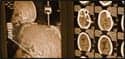
With TraumaCad, surgeons evaluate the postoperative anatomical alignment of various surgical scenarios, such as cutting, displacing, and implanting, to devise an optimal surgical plan. TraumaCad, once incorporated into the patient file, produces an accessible database of implants, including template libraries for total ankle, total knee, total hip, and total shoulder replacement.
The software can define fracture fragments, and move, rotate, and copy fragments on DICOM images and between images to reconstruct and accurately restore the anatomy on multiview images prior to templating. It also assists pediatric orthopedic surgeons in taking anatomical measurements, comparing them to normative standards, and simulating corrective procedures.
User-friendly wizards help surgeons produce a range of anatomical measurements, such as growth multiplier, acetabular index, malalignment, and spinal deformity. All measurements and evaluations are integrated into patient files.
Orthocrat will present its advanced orthopedic-templating solutions at the upcoming 94th Annual Scientific Assembly and Annual Meeting of the Radiological Society of North America from November 30 through December 5.
—Elaine Sanchez


