 |
· 3D/4D Ultrasound Lands First Buyer
· GE Fuses Ultrasound with CT, MR, and PET
3D/4D Ultrasound Lands First Buyer
With 4D imaging capability and 3D wall motion tracking, Toshiba America Medical Systems Inc’s Aplio Artida ultrasound garnered its first customer this summer.
 |
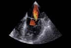
| Toshiba’s Aplio Artida can track myocardial motion and display it in 3D images. |
“Being the first to purchase the Aplio Artida is an exciting development for us and our patients,” said Richard Marcus, MD, director of the cardiovascular ultrasound laboratory at Iowa Heart Center. “Having firsthand experience with the system, I am pleased with the images it produces. Its enhanced applications, particularly the 4D imaging, will allow our facility to continue to improve the care provided to our patients.”
Designed to meet the demands of the growing cardiac 4D market, the Artida is equipped with real-time, multiplanar reformatting capabilities that allow physicians to quantify global and regional LV function, such as LV ejection fraction, volume, and severity of regurgitation. Also available to assist in surgical planning are arbitrary views of the heart that cannot be achieved through 2D imaging.
The Artida is the world’s first ultrasound that can track myocardial motion and display it in 3D images. Through 2D/3D wall motion tracking, users can obtain angle-independent, quantitative, and regional information about myocardial contraction. According to Toshiba, the ability to identify wall motion defects and heart timing will enable pacemaker setting optimization and improve Cardiac Resynchronization Therapy using pacemakers by determining whether a person will likely be a responder to CRT.
Gerard Aurigemma, MD, professor of medicine at the University of Massachusetts, said the most important job for a clinical echo lab is the evaluation of left ventricular function, and speckle tracking for this purpose is the most exciting development in cardiac ultrasound in years.
“Toshiba’s 4D tracking offers estimation of left ventricular volumes, ejection fraction, and direct measurement of global and regional myocardial function by means of speckle tracking, and provides insights that echocardiographers had not had before,” said Aurigemma, who is also director of noninvasive cardiology at the UMass Memorial Medical Center. “This type of analysis will be how the next generation of echocardiographers quantifies ventricular function.”
The ultrasound’s SmartCast Beamformer utilizes advanced digital signal processing that aims to control the shape of the ultrasound precisely and flexibly. In an effort to improve diagnostic accuracy, the system features a number of performance improvements, such as simultaneous multiple transmit focus or a doubled frame rate in color Doppler mode.
Intuitive SmartSlice functions enable physicians to cut, slice, and position the 4D volume with quickness, as a variety of prospective and retrospective volume acquisition modes provide the flexibility to acquire and store 4D volumes as raw data. Physicians can surf volumes at any time, either on the system or offline.
Furthermore, the Artida enhances diagnostic capabilities with multiple processors that run synchronously, which allows for handling of large amounts of data. Its multiple processor design employs the distributed processing power of more than 80 processor cores interconnected by a fast digital system interface. According to Toshiba, the engine is so powerful that it can process an amount of data equal to a fully loaded DVD every single second.
—Elaine Sanchez
GE Fuses Ultrasound with CT, MR, and PET
Today, manufacturers are focused on improving interventional procedures. GE just scored a home run. On September 2, the company announced the launch of its latest ultrasound system for radiology and vascular applications, the LOGIQ E9. The new system’s Volume Navigation tool enables both GPS-like marking of a patient’s anatomy during an exam and real-time fusion of imported PET, CT, or MR images.
 |
| GE’s new LOGIQ E9 is a “triple threat!” |
“We’re very excited about this,” said Terri Bresenham, vice president of diagnostic ultrasound and information technology at GE. “We’ve been working with radiologists and interventional radiologists, asking how we could help improve interventional procedures and enable a more cost-effective approach.”
The answer is real-time fusion of anatomical data with 4D information obtained by the LOGIQ E9. “The primary purpose of the system is real-time live scanning,” Bresenham explained. “So I go to the ultrasound system and recall the prior set of info I wanted—a CT, an MR, a nuclear medicine study, a prior ultrasound. I import that into the ultrasound system, and it creates a reference field around the patient. Within 60 seconds the data has been imported, and as you begin to move the probe the system presents that data from the other modality as you’re scanning live.”
As the clinician scans, anatomical information and real-time data, such as blood flow information, are integrated. “The potential for this is very exciting,” Bresenham said. “It addresses a lot of health care needs that we see. It’s low-cost, it has real-time capabilities, it’s portable, and it provides outstanding clinical information.”
The system is DICOM-compatible, so images can be imported from any standard PACS system and any scanner. “Even if you’re in an office-based setting, as long as you have access to a PACS network, you’ll be able to fuse these studies,” Bresenham said. “But a patient can also show up with a CD/ROM or a USB drive. We allow for many ways to import the prior information. There’s no restriction on where this could be used.”
Other key features of the system include its GPS-like tracking, which allows clinicians to “mark” an area of the patient’s anatomy for more precise follow-up exams. “Thyroid exams have traditionally been very time-intensive,” Bresenham said. “You’re always wondering, is this the same nodule I measured before? Now you can lay down GPS markers so that when the patient comes back, you know for certain you’re looking at the same spot.”
The system also leverages GE’s new Agile Ultrasound architecture for sonography, which uses modular mathematical models to provide more accurate measurements of how sound interacts with different tissue types within the body. “This is not to be overlooked,” Bresenham said. “When you’re sending a sound wave into someone with the current technology, you’re assuming a lot about the person you’re sending it into. With Agile, when I go to put the probe on for a liver study, the system knows I’m imaging the liver. It’s intelligent. It knows where I’m looking and what needs to be done, so the penetration capabilities are better.”
The system also features workflow enhancement in the form of Scan Assistant, a tool that allows clinicians to preprogram the actions they perform most, eliminating time-consuming keystrokes. “It’s quite literally another set of hands,” Bresenham said. “We see that the patient’s interaction with the sonographer is improved, keystrokes are reduced, and approaches to imaging are standardized.”
The LOGIQ E9 will make its debut at this year’s meeting of the Radiological Society of North America, Oak Brook, Ill. “What’s been fascinating is the number of ideas that have been borne out of seeing the system demonstrated live,” Bresenham said. “It will go well beyond the areas we initially intended. The runway for this is long and rich in ideas.”
—Cat Vasko

