 |
· Ultrasound Expertise Matters in Ovarian Cancer Diagnosis
· Siemens Ultrasound Catheter Opens to Third Parties
Ultrasound Expertise Matters in Ovarian Cancer Diagnosis
Turning to ultrasonographers with expertise rather than regular operators when diagnosing ovarian cancer results yields a significant reduction in the overall number of diagnostic procedures required and diminishes the length of inpatient hospital stays, according to a recent study published in Lancet Oncology.
 |
Researchers say that expert operators exhibit greater skill in distinguishing benign from malignant ovarian pathology. Study authors Joseph Yazbek and colleagues, from King’s College, Guy’s, and St Thomas’ hospitals in London, conclude, “This ability decreases the number of patients who are treated as potentially having ovarian cancer and aids the use of more conservative management options.”
Traditionally, ultrasound images that examine structural features, such as wall structure, blood vessels, and presence of fluid, are used to distinguish benign from malignant tumors in the uterus, ovary, or fallopian tubes. Yet, interobserver variability can substantially affect ultrasonography, as experienced operators demonstrate they are significantly more accurate in their diagnosis than those who are less experienced. With the inability to differentiate benign and malignant tumors, patients are referred for major abdominal surgery to rule out the possibility of cancer, instead of the option for minimally invasive laparoscopy to remove the benign mass or a wait-and-see approach.
With a mission to evaluate whether operator skill had a measurable impact on patient management, Yazbek and his team of researchers examined the cases of 150 patients with suspected ovarian cancer who were referred to the regional gynecological cancer center at Guy’s and St Thomas’ NHS Foundation between May 3, 2004, and February 15, 2007. These patients were randomized to level III (expert) ultrasonography—undertaken by gynecologists with a special interest in ultrasonography who had more than 10 years of experience in the procedure—or level II (routine) ultrasonography—undertaken by ultrasonographers trained in gynecological ultrasonography. For all patients, both transvaginal and transabdominal scans were performed, ensuring thorough assessment of the entire abdominal cavity.
Results revealed the number of major surgical staging procedures for presumed ovarian cancer undertaken in women by a level III ultrasonographer was 17 out of 77, or 22%. Those screened by level II ultrasonographers amounted to 27 of 73, or 37%. Additionally, there was a reduction in follow-up procedures after expert sonography with the median number of follow-up scans equaling two (range 0 to 5) in the level II group and one (range 0 to 4) in the level III group.
“This finding is likely to be the consequence of the greatly increased proportion of patients in whom a conclusive diagnosis of the nature of the adnexal tumor was possible from level III ultrasonography compared with level II ultrasonography,” according to the authors.
A histological diagnosis was provided to clinicians for 76 of 77 patients in the level III group, compared with 38 out of 73 patients in the level II group. While the total number of surgical procedures was similar between the two groups, with 35 of 73 in level II and 33 of 77 in level III, the number of minimally invasive procedures was higher for the level III group. The researchers attributed this to the significant decrease in the length of hospital stays for patients in the expert group. The median duration of hospital stay for level II patients was 6 days (range 3 to 13). For level III patients, the median duration of hospital stay was 5 days (range 1 to 9).
The effect of expert scanning might have been greater if it had been used in the primary assessment of ovarian pathology, the authors added. “Increased confidence in the diagnosis of benign ovarian lesions is likely to decrease the need for additional diagnostic tests, such as MRI or serum CA-125 concentration, and also decreases the number of referrals to regional cancer centers,” the researchers wrote.
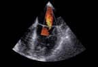
Siemens Ultrasound Catheter Opens to Third Parties
There’s exciting news for both the industry and heart patients. Malvern, Pa-based Siemens Healthcare recently announced an extension of its strategic alliance with Biosense Webster Inc, Diamond Bar, Calif, that opens up the ACUSON AcuNav?intracardiac?ultrasound catheter for use on third-party ultrasound systems.
 |
“With the clinical benefits that can come from the use of this imaging technology, it’s?easy to see?that you’d want to extend it to all patient populations,” said Louise Kruz, global cardiology segment manager, Ultrasound,?Siemens Medical Solutions USA Inc. “No one imaging?company?dominates?all areas of clinical imaging globally. So opening it up to other vendors allows this clinical benefit to reach more patients.”
In 2006, the two companies announced a deal that provided Biosense Webster with exclusive worldwide rights to distribute Siemens’ ACUSON AcuNav ultrasound catheters.?
Imaging with the AcuNav catheter?enables electrophysiologists and interventional cardiologists to view real-time diagnostic ultrasound images and Doppler blood flow information from within the heart. Kruz said the catheter is unique in that it offers full ultrasound imaging with a large imaging field of view at the end of a catheter tip, and therefore it covers all of the traditional ultrasound imaging modes?and quantification applications?that one would expect from a cardiology transducer.
Kruz also mentioned that the ultrasound catheter is a well-accepted?alternative to x-ray-guided radiofrequency ablation. “Physicians?get more accurate information about the tissue that they are trying to?treat,” she said. “Also, EP procedures can take hours,?so monitoring?with ultrasound?reduces exposure to the associated radiation?for the patient and the medical personnel involved.”
In terms of device placement, the AcuNav catheter allows health care providers to see exactly what they are doing, Kruz added. It allows for patient monitoring with immediate feedback, and therefore, clinicians can adjust plans as they conduct the procedure.
“Oftentimes, when you are dealing with?providing treatments or making?corrections?within the heart,?general?anesthesia is required,” Kruz said. “But many of the?AcuNav-guided?procedures can use a local?for catheter insertion, and the patient doesn’t have to undergo the risk of general?anesthesia. This allows?treatment options?or interventional procedures to be?extended?to?a wider range of patients.”
Kruz said Siemens is in the process of introducing the?AcuNav?catheter for use with one of GE’s laptop systems. She pointed out that within the OR and treatment room environments,?a laptop?ultrasound system with?its easy?portability is particularly advantageous.
Siemens already features products, released and in use around the world, that are open to images of other vendors:?syngo Velocity Vector Imaging, which allows for visualization of the motion of the?heart walls, and Auto Left Heart, which uses progressive pattern recognition for expert-like measurements of ejection fraction and volumes of the left ventricle and?left?atrium.