
Low-Tech Telemammography System Based on JPEG 2000 Reduces Recalls
BioLucent Releases New MammoPad for Gamma Imaging
Intraoperative Radiography: Did We Get All the Cancer?
Product Showcase: New Version of Mammography CAD Solution Now Available
Low-Tech Telemammography System Based on JPEG 2000 Reduces Recalls
By Judith Gunn Bronson, MS
Fewer and fewer American women have easy access to mammography, as the number of sites providing the service continues to shrink. As a result, and because parts of the country have always been poorly served, considerable work has been done with telemammography. However, telemammography has faced a literally huge problem: the size of full-field digital mammograms, which are 35 megabytes (MB) to 55MB per image.
 |
The assumption has been that a high-capacity system is necessary. The system would require a broadband Internet connection; a dedicated high-speed network;1 or satellite communication, such as the Satellite Telemammography Project involving NASA’s Lewis Research Center, Cleveland. However, in a laboratory simulation, a team of researchers from the University of Pittsburgh and the Magee-Womens Hospital of the University of Pittsburgh Medical Center recently demonstrated the use of low-tech telemammography for the purpose of reducing patient recalls.2 The system has now been in clinical use for more than 2 years.
The imaging site is equipped with a personal computer operating on a Microsoft Windows 2000 Workstation. It is connected to both a Lumiscan high-resolution laser film digitizer from Kodak Health Group, Rochester, NY, and a Scanjet 5470c document scanner from Hewlett-Packard, Palo Alto, Calif.
When, after reviewing the films, a technologist finds something that is suspect, the films—along with any previous images of the same patient—are digitized with a pixel size of 50 ?m. A CAD protocol is applied, after which the images are reduced to a more manageable size, first by cropping them to remove all the areas that do not depict tissue and then by 75:1 compression with the lossy 9/7 transform wavelet-based JPEG 2000 method. Medical reports on the patient are captured with the document scanner. The material is now ready to be sent to a central site for review by radiologists using either a 56K modem over a standard telephone line or by an Ethernet network.
For this step, the technologist constructs a chat message using pull-down menus that specify which breast requires attention, which view is being sent, what the technologist sees that requires radiologist attention (such as asymmetry or a palpable lump), any information about previous mammograms (such as new finding or moderate change), and what additional procedure might be necessary (such as additional views). The system provides a generic image of both breasts for the technologist to mark the site of the remarkable finding.
The central site is equipped with a dual-processor telemammography workstation, the Athlon MP from Advanced Micro Devices, Sunnyvale, Calif, which is running on a Windows 2000 Server. This workstation has three 2,048 x 2,560 8-bit grayscale portrait monitors and two Dome C5i flat-panel monitors from Planar Systems, Beaverton, Ore, to display the images, as well as one DS5100P CRT monitor from Clinton Electronics, Rockford, Ill, to display the message. The images coming in from the remote site are unencrypted, decompressed, and reconstructed. The radiologist can display multiple images simultaneously, obtain magnified views of chosen areas, view the CAD findings as an overlay, and read the message from the technologist. If desired, images can be printed to film. After reviewing the images, the radiologist sends a text message back to the technologist with directions. The entire process can be completed while the patient waits.
The trial run of this system produced impressive results. A series of four test cases, each consisting of four images, could be sent in less than 7 minutes. Radiologists were not able to distinguish images that had and had not been compressed with the JPEG algorithm. The system, now in clinical use, has been employed in more than 2,400 cases, and it appears that recalls can be reduced by as much as 50% without the performance of an unreasonable number of “unnecessary” secondary procedures.
Judith Gunn Bronson, MS, is a contributing writer for Medical Imaging. For more information, contact .
References
- Sheybani EO, Sankar R. ATMTN: a telemammography network architecture. IEEE Trans Biomed Eng. 2002;49:1438?1443. Available at: ieeexplore.ieee.org/xpl/freeabs_all.jsp?isnumber=25972&arnumber=1159136&count=12&index=6. Accessed October 13, 2006.
- Leader JK, Hakim CM, Ganott MA, et al. A multisite telemammography system for remote management of screening mammography: an assessment of technical, operational, and clinical issues. J Digital Imaging. 2006;19:216?225. Available at: springerlink.com/content/qh2586407586043h/. Accessed October 13, 2006.
BioLucent Releases New MammoPad for Gamma Imaging
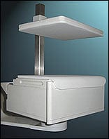 |
| The new MammoPad for gamma imaging increases patient warmth and comfort during the 6- to 10-minute adjunct procedure. |
BioLucent Inc, Aliso Viejo, Calif, has announced FDA clearance of a new version of its MammoPad breast cushion, designed specifically for use with the 6800 Gamma Camera from Dilon Technologies LLC, Newport News, Va. The 6800 is used for breast-specific gamma imaging, an adjunct diagnostic procedure to mammography wherein the breast is immobilized for 6 to 10 minutes per view.
The radiolucent foam cushion does not interfere with imaging of the breast, but it does line the detector and the shield during mammographic procedures, providing additional patient warmth and comfort. The pad’s gripping effect also helps to stabilize the breast during long-duration imaging. Clinical studies in the United States and Sweden have shown that three out of four women experience a 50% decrease in discomfort when a pad is used during mammography, according to the company.
MammoPad cushions are intended for one use only, and the foam used is biostable and recyclable. The pads can be used on any mammography machine, from analog to full-field digital. Benefits include improvement in image acquisition from improved patient relaxation and minimized patient movement. For more information, visit www.mammopad.com.
Intraoperative Radiography: Did We Get All the Cancer?
By Judith Gunn Bronson, MS
That is the question always asked during an image-guided excision of a nonpalpable breast cancer—that is, at least half of all such cancers. The classic means of finding the answer has been to send the excised tissue over to the radiology department, where two orthogonal views are obtained by a technologist and examined by a radiologist. Meanwhile, the operative team waits and the patient remains anesthetized until the radiologist calls the surgeons to say that the lesion has (or has not) been removed and the surgical margins do (or do not) appear clear.
A team from the University of Washington, Seattle, and the Bellingham Breast Center, Bellingham, Wash, thought there must be a better, faster way, and it appears they’re right. The team tested the value of the Faxitron MX-20 digital system from Faxitron X-ray Corp, Wheeling, Ill, in 150 consecutive patients, comparing it with the standard radiology-department films. (Patients were charged for only one set of films.) The MX-20, which has a footprint of 17 x 18.5 inches and includes a dedicated workstation with a 19-inch monitor, can be installed in or just outside the operating room. It captures digital images that are displayed within 15 seconds. A member of the operating team transfers the resected tissue to the instrument and triggers image acquisition, which is obtained with automatic exposure control. Moreover, no radiation precautions are needed with the shielded equipment. The surgeon views the high-resolution (10 linepairs/mm) images, which may be obtained from multiple angles if necessary. Magnification up to 5 times is built in, making microcalcifications as small as 10 ?m visible. Films can be printed if required. The equipment can be applied to smaller lesions, such as core biopsies, as well as resected tissue, to ensure that the desired tissue has been harvested.
In the Washington clinical trial—highlighted at the Seventh Annual Meeting of the American Society of Breast Surgeons in Baltimore in April 2006 and published in the American Journal of Surgery 1—the surgical and nursing team found that the traditional and Faxitron images were equally accurate in depicting the breast lesions and surgical margins. Among the patients with positive or questionable margins, 58% underwent further excision immediately on the basis of the images, and 47% achieved negative margins. No significant difference in the accuracy of the interpretation by the surgeons and radiologists was detected. Overall, use of the intraoperative system alone would have shortened the procedure by an average of 19 minutes, and no radiologist or technologist would have been needed. “Surgical review of prompt intraoperative images speeds surgery without loss of accuracy,” the team concluded.
How is the Faxitron being used now? “At the moment, for medicolegal reasons, we are still using the radiology department to overread the films,” according to Cary S. Kaufman, MD, FACS, lead author of the study. “There also is a question regarding who will write the final report for the chart. However, we routinely use intraoperative ultrasonography for localization and make surgical decisions about the need for further resection prior to any reading by the radiologists on the basis of the surgeon’s reading of the Faxitron images in the operating room.
“There is a difference between diagnosing a lesion on mammography and seeing that lesion on a specimen mammogram after you have removed it,” Kaufman continues. “We are not providing initial image diagnosis, and everyone should recognize that. The radiologist provides the image diagnosis; we simply identify the lesion again, which is easier than finding it in the first place. The result saves time without losing quality.”
Kaufman’s advice is to use the Faxitron routinely in the operating room. “You will see how much time it saves and will not return to waiting for the films in radiology,” he says. “A PACS will allow the radiologist to review the films promptly. With double reading, we may not be saving money yet, although I suspect the reading in the operating room is cost effective. Certainly, we are saving time for both the patient and the surgeon.”
Judith Gunn Bronson, MS, is a contributing writer for Medical Imaging. For more information, contact .
Reference
- Kaufman CS, Bachman BA, Jacobson L, Kaufman LB, Mahon C, Gambrell L. Intraoperative digital specimen mammography: prompt image review speeds surgery. Am J Surg. 2006;192:513?515. Available at: www.ajsfulltextonline.com/article/PIIS0002961006004296/abstract. Accessed November 9, 2006.
Product Showcase: New Version of Mammography CAD Solution Now Available
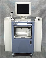 |
| CAD v7.2 from iCAD offers reduced false markings for improved diagnostic accuracy. |
iCAD Inc, Nashua, NH, has released version 7.2 of its mammography CAD solution. The new version will ship with all of iCAD’s SecondLook 300 and SecondLook 700 systems, offering the ability to automatically detect potentially cancerous areas based on algorithms.
“Though mammography has come a long way, there is still a need for increased sensitivity,” Rachel Brem, MD, director of breast imaging and intervention at the George Washington University Medical Center, Washington, DC, said in a press release. “iCAD’s latest software version has improved sensitivity to subtle cancers with fewer false positives and far more understandable indications. Tests at my facility showed a significant improvement in mass detection.”
iCAD detects areas of concern using advanced pattern-recognition techniques, then analyzes them with an intelligent algorithm, thereby identifying subtle lesions that could otherwise be overlooked. In clinical studies, the system has been shown to find up to 72% of cancers that had been missed on the previous mammogram exam. V7.2 offers reduced false markings of vascular calcifications and lymph nodes as well as reduced false markings due to linear structures, film noise, or film grain. For more information, visit www.icadmed.com.


