
Outsourced Digitizing Service Eases Transition to Digital Mammography
Running the Numbers
Product Showcase: Mammography Digitizer Saves Space, Time
KB-CAD System Provides “Search Engine” for Mammography
Product Showcase: Mobile Stitching Cassette Holder Designed for Long-Length CR Images
Product Showcase: Skin Markers Geared Toward Women With Sensitive Skin
Outsourced Digitizing Service Eases Transition to Digital Mammography
 |
| Outsourced digitizing can help to ease the transition from analog to digital mammography. |
Results from the American College of Radiology Imaging Network Digital Mammographic Imaging Screening Trial (ACRIN DMIST), published in 2005, suggest that digital mammography might be better than analog for detecting breast cancer in certain cases. But the conversion from analog to digital can be complicated, time-consuming, and costly. A new service from Digi-Solve LLC (Ponte Vedra Beach, Fla) aims to facilitate this transition by offering an outsource service that digitizes archived images.
“The price of digitizers is climbing,” says Digi-Solve President John DeBiase. “On top of that, there’s the labor expense and something we think of as a ?hassle’ expense—think about feeding thousands of films all day long into a machine, and that’s all you do.”
DeBiase’s alternative is for a facility to ship its old films to Digi-Solve, where college students, retirees, and disabled persons—all paid on a per diem basis—will feed the films into digitizers from VIDAR Systems Corp (Herndon, Va). “We’re outfitted to be able to handle 20 machines going 16 hours a day,” DeBiase explains. The data will then be processed using Digi-Solve’s breast-specific DigiScan software, after which the images are sent back to the facility using a secure T3 Internet connection. Turnaround time is just 1 to 2 days.
The outsourced solution can save facilities money, DeBiase says, as well as improve workflow. “A lot of departments load a multiviewer,” he explains. “Then, the radiologist comes in and deals with the light problems in the room and the contrast differences between the multiviewer and the workstation. After the multiviewer is all used up, the radiologist must stop reading. With the shortage of radiologists reading mammo today, that creates a problem. With our service, facilities can eliminate the space constraints and limitations of the multiviewer, so a radiologist can keep being productive.”
DeBiase estimates that the outsourced solution could save facilities 20% to 25% compared with in-house digitizing. “What we recommend to folks is that they give us their schedule 2 weeks out, and we digitize all their patients for the week, whether it’s 1, 2, or 3 years prior—the customer’s choice. Then, we digitize all of their films and send the files to either their workstations, their PACS, or an outsourced provider,” he says. “That’s the most cost-effective for them.”
—C. Vasko
Running the Numbers
54% more likely to develop breast cancer were women identified as BRCA 1 or 2 carriers who had ever had a chest x-ray than women who had never undergone the procedure, according to a recent study.1 The International BRCA 1/2 Carrier Cohort Study, conducted by a consortium of European cancer centers, analyzed questionnaires from 1,600 women who carry BRCA 1/2 mutations. Researchers also found that women who were exposed to x-rays before age 20 had a 2.5-fold increased risk of developing the disease before age 40, compared with women who had never been exposed. “Since BRCA proteins are integral in repairing damage to breast cells, we hypothesized that women with BRCA 1/2 mutations would be less able to repair damage caused to DNA by ionizing radiation,” said David E. Goldgar, PhD, a lead author of the study who was the chief of the Genetic Epidemiology Group at the International Agency for Research on Cancer (Lyon, France) at the time the research was conducted. “Our findings support this hypothesis and stress the need for prospective studies.”
Reference
- Andrieu N, Easton DF, Chang-Claude J, et al. Effect of chest x-rays on the risk of breast cancer among BRCA 1/2 mutation carriers in the international BRCA 1/2 carrier cohort study: a report from the EMBRACE, GENEPSO, GEO-HEBON, and IBCCS Collaborators’ Group. J Clin Onc. 2006;24:3361?3366. Available at: http://www.jco.org/cgi/content/abstract/24/21/3361. Accessed September 8, 2006.
Product Showcase: Mammography Digitizer Saves Space, Time
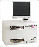 |
| Although the 2908 MammoPro digitizing system from Array is controlled by a touch screen, it can be enhanced with a keyboard and mouse. |
A new digitizer from Array Corp USA (Hampton, NH) is now available. The 2908 MammoPro system offers a tabletop digitizing system controlled by a touch screen, eliminating the need for a separate PC controller; the system is also compact and self-contained, measuring 26 x 33 x 16 (WHD).
The 2908 MammoPro features a 50-?m noise-reducing system, which can penetrate films up to density OD4 at a speed of less than 30 seconds for an 18- x 24-inch film. It also boasts the same engine as the ImageChecker DMax computer-aided detection (CAD) system for digital mammography from R2 Technology Inc, a Hologic Co (Sunnyvale, Calif). The system is compatible with all major DICOM-based CD-burning solutions, and the touch-screen interface can be enhanced with an optional keyboard and mouse.
“The Array 2908 MammoPro complements the emerging digital marketplace by allowing users to capture relevant priors in a productive, high-quality, ergonomic environment,” Array President Thomas Nardozzi said in a press release. “We believe it to be the world’s first fully self-contained digitization system for customers demanding speed, flexibility, and superior imaging quality.”
For more information, visit www.arrayusa.com.
KB-CAD System Provides “Search Engine” for Mammography
A new system developed by medical physicists at Duke University (Durham, NC) can speed up the process of computer-aided detection (CAD) using a search-engine-like query of a mammogram database. The system, known as knowledge-based computer-assisted detection (KB-CAD), works by comparing mammograms to images of known breast cancer using the principles of information theory for better speed and efficiency.
“The user starts by clicking on an image location that appears suspicious,” explains Georgia Tourassi, PhD, associate research professor in the department of radiology at the Duke University School of Medicine, who presented the system at the 48th Annual Meeting of the American Association of Physicists in Medicine (AAPM) in Orlando, Fla. “The system compares the image neighborhood around the query location with images stored in the knowledge database. Our KB-CAD system is intended as an interactive computer aid to help the user analyze any mammographic location he or she wants.”
KB-CAD differs from ordinary CAD, however, in that it maximizes system performance by comparing the unknown case with the most informative stored cases. Tradi-tional CAD systems are weighed down with comparing the mammogram in question to every image in the database; however, KB-CAD indexes its cases based on image entropy.
“Entropy is used to quantify image complexity by looking at the distribution of the gray level values of the image pixels,” Tourassi explains. “Images with greatly varied distributions of image pixel values have higher entropy than images with more uniform distributions. The normal breast parenchyma can be as complex as abnormal tissue in mammograms, often resulting in similar entropies.
“In our preliminary studies,” she continues, “we observed that knowledge cases with higher entropy—in other words, higher complexity—are more useful to the system in terms of making better decisions. This result suggests that instead of blindly depositing mammograms in the knowledge database, we may need to consider a more selective deposit scheme where only complex mammograms with higher entropy are deposited.”
For their pilot study, Duke researchers debuted KB-CAD on a database of 2,300 images. Each sample image was compared to only the 600 most informative stored cases as determined by image entropy, which cut analysis time to less than 3 seconds per search. “Entropy calculations are very fast,” Tourassi says. “The calculation of mutual information is computationally very expensive. Thus, by following the entropy-based indexing scheme, we can substantially cut down on the computational demands of the KB-CAD system without sacrificing diagnostic performance.” Tourassi says there are no particular requirements for the technology used to implement the system.
The researchers have now turned their attention to optimizing the system parameters, studying the clinical impact of the interactive paradigm, and developing a system that will easily translate across a variety of platforms.
—C. Vasko
Product Showcase: Mobile Stitching Cassette Holder Designed for Long-Length CR Images
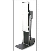 |
| The Mobile Stitching Cassette Holder is a space-saving solution for viewing long-length images. |
The new 14- x 51-inch Mobile Stitching Cassette Holder from Konica Minolta Medical Imaging Inc (Wayne, NJ) addresses the issue of limited wall space in radiology departments and offers a display solution for long-length images attained by the company’s REGIUS Xpress CR, IQue CR, and IQue SE.
The unit, which can be attached to and removed from the wall at will, includes a built-in grid with a ratio of 10:1, a line pair per inch of 178, and a focal distance of 60 to 90 inches; it is height-adjustable and has a traveling distance of 38 inches. Image-stitching software is necessary for use.
“The Mobile Stitching Cassette Holder is [ideal] for radiology departments that do not wish to permanently affix a mount for stitching to their x-ray room walls,” Eunice Lin, marketing and product planning manager at Konica Minolta Medical Imaging, said in a press release.
Product Showcase: Skin Markers Geared Toward Women With Sensitive Skin
 |
| Beekley?s Soft ?n? Stretchy SPOTS mark scars, moles, and nipples during mammography exams. |
Beekley Corp (Bristol, Conn) recently introduced the latest addition to its line of mammography skin markers. The new Soft ‘n’ Stretchy SPOTS, which mark scars, moles, and nipples, are designed to stick better and peel away with less resistance, resulting in a more comfortable patient experience.
The new markers are intended to reduce the need for retakes by sticking better during positioning and compression; the material also bends and expands with the patient’s breast during compression, giving technologists the opportunity to focus more on completing the scan with all possible expedience. When the exam is finished, two tabs are pulled in opposite directions, allowing the sticker to be lifted away quickly and painlessly.
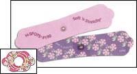 |
Soft ‘n’ Stretchy SPOTS come in four shapes for unmistakable images on film, and they are available in two designs: pastel pink or floral. For more information or to request samples, visit www.beekley.com.

