Breast imaging providers offer smart strategies for improving mammography workflow and your bottom line.

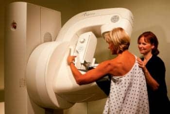
Fujifilm?s recently FDA-cleared Aspire? HD has the potential to increase throughput and diagnostic confidence.
In 2007, Sherry Gage, RTRM, was asked to spearhead efforts to create a new women’s breast center in Phoenix. Technologically, the new center was going to be state-of-the-art, even eventually offering breast tomosynthesis, or 3D mammography.
But, even beyond revolutionary new technologies, the John C. Lincoln Breast Health and Research Center was going to create an optimal workflow environment so that the center would not only provide a relaxing, stress-free environment for its patients, but would also, in Gage’s words, “make money.”
But making money, says Gage, is not an easy thing to do when it comes to mammography. “The technology is expensive, there are lots of regulations to follow, and not much whatsoever in the way of reimbursement,” she said, which means that optimizing workflow would be critical in making the new center financially successful.
While optimizing workflow was a primary goal, it was important to ensure that patients wouldn’t experience something approaching a “cattle call,” Gage said, “Which is always a concern when you’re trying to increase workflow.”
So when the new center finally opened in May 2009, a lot of money had been spent creating a facility that was almost spa-like, says Gage, with one mammography room and plenty of room for growth.
And the breast center would need the extra space. According to Gage, the center’s popularity has skyrocketed in the first 2 years of its existence. It now performs about 2,500 exams a month, compared to 400 when it opened. The breast center’s success came so fast that it opened up two more mammography rooms within its first 6 months.
In order to make those numbers manageable from a workflow perspective, Lincoln had to maximize its radiologic technologists’ time doing what they do best—performing exams.
“The people who have the biggest impact on workflow are the technologists,” said Gage. “And tech staffing is expensive, at least in Arizona.” The problem, she says, is that technologists customarily spend about 75% of their time doing paperwork in order to meet requirements like those of the Mammography Quality Standards Act, or the American College of Radiology accreditation program.
“So the first thing we did was remove the paperwork from the techs—everything except the patient history form,” Gage said. The breast center did it by hiring technologist assistants who are tasked with entering all of the paperwork into the center’s RIS and mammography information system.
The center puts two radiological technologists in each mammography room, and while one greets a patient and takes her history, the other performs a procedure on another patient. “It’s very important that the room stay busy,” said Gage, “but this system also removes the ‘cattle call’ appearance.”
The system not only improves workflow and the patient’s experience, it also has a positive impact on quality. The technologist is taking the time to sit down with the patient and methodically take down her history, which means that form is going to be more accurate.

The team from the John C. Lincoln Breast Health and Research Center including Michael Alpern, MD, Sherry Gage, RTRM, and Linda Greer, MD, Medical Director of the Breast Health and Research Center, with their Hologic Selenia Dimensions breast tomosynthesis unit.
Stressing Patient Comfort
Making the mammography process more patient friendly can “come at a cost regarding volume,” said Ayala Rosenbaum, MD, a radiologist at Rosetta Radiology in New York City. “But we believe it’s the right way to practice medicine. We never want to forget that each image represents a human being.”
According to Rosenbaum, patients coming in strictly for mammograms are in and out of Rosetta’s doors in 30 minutes. The key, she says, is ensuring that the practice has multiple staff members tracking each patient.
When a patient arrives at Rosetta, administrative staff immediately start tracking patients through the practice’s RIS. Once the patient checks in, she shows up on the work list of every modality for every test that patient is scheduled to take. If at any time that patient’s name is highlighted in red, it means the patient has been waiting too long and needs to be seen.
Patients are triaged to different floors depending on the exam, and on each floor a receptionist greets the patient, checks to see what technologist that patient is scheduled to see, escorts the patient to the dressing room, and watches over her until she is seen.
Technologists are always aware of whom they’re supposed to be seeing and when they’re supposed to see them. And when exams are completed, the results are presented to the physician. “So we have three people watching the patient,” Rosenbaum said. “The physician, a technologist performing one test, and a technologist waiting to perform the next one.”
And once the patient has been handed over to the physician, there should be a minimal wait for the patient to get the exam results. “It really doesn’t take a lot of time and effort to give a patient her results on the same day,” Rosenbaum said. “It shouldn’t take more than a minute to tell a patient you’re fine and we’ll see you next year.”
Because patients get results so quickly, recalls are handled on the spot. “So we don’t have to review cases weeks later to see why the patient has been recalled and what information needs to be retrieved,” said Rosenbaum. “So that saves time in the long run.”
Despite the fact that Rosetta’s approach comes at a cost when it comes to volume, there are positives financially.
“Patients know they are going to be treated like human beings, and this helps distinguish us from other practices,” said Rosenbaum. “Mammography serves as a loss leader because we’re developing loyal patients who will come back to us for any kind of imaging they need.”

Chinese Hospital?s Chief of Radiology Roger Eng, MD, MPH, can read digital mammography images along with general radiology exams on a Carestream PACS workstation.
Digital Impact
Up until 2007 the radiology department at Chinese Hospital relied literally on paper and film. “We had no RIS or PACS,” said Roger Eng, MD, MPH, chief of radiology at the hospital. “We had pads of paper and schedule books, and that was our scheduling systems.”
Wait times for mammography appointments ran into months, and on the day of an appointment a patient faced an excruciatingly long wait to be seen.
The department’s challenges went beyond archaic technology. “If you know anything about Chinatown in San Francisco, you know that space is at a premium,” said Eng. “We are space challenged—our footprint is about half the size of a typical radiology department.”
The waiting room was so small, Eng says, that patients had to wait out in a hallway and were supplied with pagers, much like you would receive if you were waiting for a table at a restaurant. “We used to have patients yelling out in the hallway because they were waiting so long,” Eng said.
That all began to change with a move to digital, but not without some growing pains, says Eng.
The problem, he says, is that the department initially believed it could improve workflow by adding a digital mammography room and keeping an existing analog room.
“We found that by having two different technologies, we were having trouble just doing 20 exams a day,” said Eng. “The problem was that we had two different workflows, one digital and one analog.” This had a negative impact on workflow all the way from patient check-in to technologists performing the exam and radiologists reading the studies.
So the department made the decision to decommission the analog room, expand the waiting area, and go with one completely digital mammography room.
“Two technologists are able to tag team in that room,” said Eng. “And we’ve been able to optimize workflow by getting rid of analog. Now we can do 30 exams a day and we haven’t even met capacity.”

Sherry Gage, RTRM, Breast Center Director, John C. Lincoln Breast Health and Research Center
New Technologies and Workflow
In April 2011, the John C. Lincoln Breast Health and Research Center became one of the first facilities in the country to begin using the Selenia Dimensions 3D digital mammography system, developed by Hologic.
The technology, called tomosynthesis, provides individual images of thin layers of breast tissue, which yields much clearer images, particularly of dense breast tissue.
“We knew it was going to be fantastic,” said Gage, “so we had one unit installed.” According to Gage, the idea was to offer the technology to women who ranged from having normal to a high amount of dense breast tissue.
The center heavily publicized the new technology in television spots and with education events, “and when it hit, it was not at all what we expected,” said Gage. “By the end of April, we had women driving in from Utah and flying in from New Mexico and California to use it.”
Before long, women who didn’t even have dense breast tissue, but instead had fatty tissue with a family history of breast cancer, were demanding the technology. And if they demanded it, the center provided it.
Of the center’s three examination rooms, one was dedicated to tomosynthesis. “Now we had about 70% of our patients who wanted tomosynthesis,” Gage said. “I had to do 70% of our exams in one room and it killed our workflow because the response to the new technology was so amazing.”
Even more amazing, Gage says, was the fact that within the first 30 days of the time the new technology was used, tomosynthesis found a four-millimeter stellate mass in the breast of a women with fatty tissue. And this hadn’t shown up on a regular digital scan.
“This had more of a negative impact on our workflow,” said Gage. “Since we had found something in a breast with fatty tissue, we felt that we had to offer tomosynthesis to everybody. Probably 85% of the patients wanted it and at times we had a 2-hour wait for mammography. It was horrible.”
The answer was simple, if expensive. The center bought another tomosynthesis unit. “Now we’re doing fantastic with workflow,” Gage said.
Tomosynthesis doesn’t take any longer than traditional digital scans, and there are other workflow benefits as well. Recall rates have gone down, and tomosynthesis also minimizes the number of views of the breast needed, even in recalls. “So we’re not spending extra time looking at different areas of the breast or calling patients back,” said Gage.
More importantly, tomosynthesis has found 10 cancers that would have been missed with traditional digital scans, Gage says, all by using a technology that requires shooting fewer images, emits a lower radiation dose, and causes less anxiety for the patient.
And while workflow has increased, there was a lesson learned, Gage says. “One machine can have a negative impact on workflow because of the response to technology,” she said. “But it can be helpful—as long as you put in more than one!”
Michael Bassett is a contributing writer for Axis Imaging News.