In a case report published in the British Medical Journal, Italian researchers describe a total hip arthroplasty (THA) in a 75-year-old woman that used 3D modeling software and 3D printing to improve the preoperative surgical planning.
CT scan is considered the gold standard to define the fracture pattern; however, the presence of the prosthetic implants in situ limits the full view of the articular surface and bone loss. A three-dimensional (3D) modelling software allows precise tridimensional reconstructions of the bony surface, virtually removing the metallic implants through DICOM image segmentation. We highlight the case of a periprosthetic acetabular fracture around THA which occurred to a 75-year-old woman, in which a 3D modelling software was used to improve the assessment of fracture morphology and bone quality. Moreover, the 3D images were printed in a real-life size model and were used for preoperative implant templating, sizing and surgical simulation.
Read more from 3DPrint.com and find the study in BMJ.
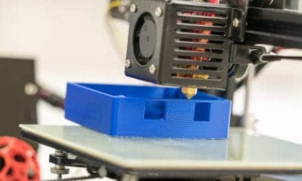
Featured image: 3D printed, real-size, plastic model of the entire pelvis. (b,c) Particular of the fracture of the medial wall and posterior column. References:Radiologia, ASL CAGLIARI, P.O. Marino – Cagliari/IT