The University of California’s two medical centers, UCSF and UCLA, and their nuclear medicine teams have obtained approval from the U.S. FDA to offer a new imaging technique for prostate cancer that locates cancer lesions in the pelvic area and other parts of the body to which the tumors have migrated.
Known as prostate-specific membrane antigen PET imaging, or PSMA PET, the technique uses positron emission tomography in conjunction with a PET-sensitive drug that is highly effective in detecting prostate cancer throughout the body so that it can be better and more selectively treated. The PSMA PET scan also identifies cancer that is often missed by current standard-of-care imaging techniques.
“UCLA and UCSF researchers studied PSMA PET to provide a more effective imaging test for men who have prostate cancer,” says Jeremie Calais, MD, MSc, an assistant professor at the David Geffen School of Medicine at UCLA. “Because the PSMA PET scan has proven to be more effective in locating these tumors, it should become the new standard of care for men who have prostate cancer, for initial staging or localization of recurrence.”
A clinical trial conducted by the UCSF and UCLA research teams on the effectiveness of PSMA PET proved pivotal in garnering FDA approval for the technique at both universities. The PSMA drug used in the technique was developed outside the U.S. by the University of Heidelberg.
“It is rare for academic institutions to obtain FDA approval of a drug, and this unique collaboration has led to what is one of the first co-approvals of a drug at two institutions,” says Thomas Hope, an associate professor at UCSF. “We hope that this first step will lead to a more widespread availability of this imaging test to men with prostate cancer throughout the country.”
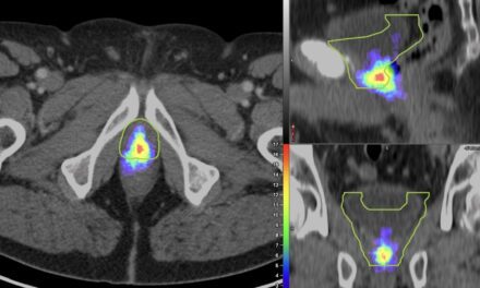
How It Works
For men who are initially diagnosed with prostate cancer or who were previously treated but who have experienced a recurrence, a critical first step is to understand the extent of the cancer. Physicians use medical imaging to locate cancer cells so they can be treated.
PSMA PET works using a radioactive tracer drug called 68Ga-PSMA-11, which is injected into the body and attaches to proteins known as prostate-specific membrane antigens. Because prostate cancer tumors overexpress these proteins on their surface, the tracer enables physicians to pinpoint their location.
The current standard of care in prostate imaging is a technique called fluciclovine PET, which involves injecting patients with fluciclovine, a synthetic radioactive amino acid. Since prostate cancers consume more amino acids than normal prostate cells, the tumors accumulate large amounts of the synthetic tracer, making them easier to detect during scans.
In their research comparing PSMA PET and fluciclovine PET, the UCLA and UCSF research teams found that imaging with PSMA PET was able to detect significantly more prostate lesions than fluciclovine PET in men who had undergone a radical prostatectomy but had experienced a recurrence of their cancer.
Their findings indicate that PSMA PET should be strongly considered both before initial treatment in men with high-risk cancers and in cases of cancer recurrence after surgery or radiation to provide more precise care. The PSMA tracer also can be used in conjunction with CT or MRI scans.
“‘Game changer’ is almost an understatement for how prostate cancer patient care could be improved by this technique,” says Jonathan W. Simons, CEO of the Prostate Cancer Foundation. “After investing more than $26 million in research on PSMA over many years, we are honored to congratulate the research teams at UCSF and UCLA on their milestone achievement.”