Researchers developed a technique combining artificial intelligence and mathematical models to produce one image per second during dynamic scans.
Researchers from the Technion-Israel Institute of Technology and the United States have developed a method to accelerate and improve magnetic resonance imaging (MRI) scans by combining artificial intelligence with mathematical models.
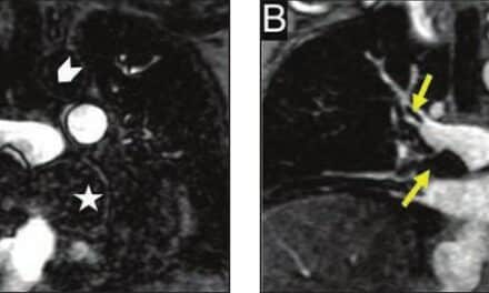
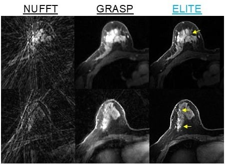
The study, published in Nature Communications, focuses on enhancing dynamic MRI for breast cancer imaging. The new method, called ELITE, enables dynamic scans with increased speed by combining artificial intelligence with models that identify structural and functional patterns in different tissues.
Dynamic MRI is a critical technology in breast cancer diagnosis, particularly for screening populations at high risk. It is characterized by sensitivity and accuracy of more than 90%, compared to approximately 50% to 60% for ultrasound and mammography combined. However, producing detailed images usually requires longer scan times, which makes it difficult to track the flow of contrast material through tissue in real time. Traditional exams typically provide one image every one to two minutes.
The researchers bridged this gap by using a deep neural network trained to remove noise and distortions, alongside the reconstruction of missing information from undersampled measurements. This process results in the generation of one image per second.
The ability to track the movement of a contrast agent almost continuously allows physicians to identify small tumors more accurately, distinguish between benign and malignant tumors, and characterize biological tumor properties such as blood flow and vascular permeability. In a study involving 54 patients, the researchers achieved improved tumor visibility compared to existing methods, high image quality, and high diagnostic sensitivity.
The study focuses on dynamic MRI, a critical technology in breast cancer diagnosis, according to Eddy Solomon, PhD, the paper’s lead author and a researcher at the Technion Faculty of Biomedical Engineering, in a release. Solomon notes that shortening scan times is expected to increase the number of women who can be scanned using a given MRI machine.
While the method was tested specifically on breast cancer imaging, the researchers demonstrated that the technique may also be useful for brain, head, and neck imaging. The method has the potential to improve other imaging platforms, paving the way for systems that enable fast and personalized imaging while providing physicians with real-time biological insights.
The study included researchers from Weill Cornell Medical College and the New York University Center for Advanced Imaging Innovation and Research. The work was supported by grants from the National Institutes of Health and Radiological Society of North America Research.
Photo caption: The new method, called ELITE, enables dynamic scans with increased speed by combining artificial intelligence with models that identify structural and functional patterns in different tissues.
Photo credit: Technion-Israel Institute of Technology