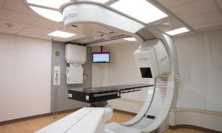
A portable point-of-care system developed at Washington University in St Louis uses a robotic arm to image organs at the bedside.
Researchers have developed a portable, point-of-care positron emission tomography (PET) technology capable of imaging any organ to guide interventional procedures in real time. The bedside technology, presented at the Society of Nuclear Medicine and Molecular Imaging 2026 Annual Meeting, aims to provide a cost-effective method for hospitals to perform biopsies, tumor ablations, and other procedures in constrained clinical environments.
Current interventional procedures, which are minimally invasive, image-guided techniques used to diagnose or treat conditions, rely mainly on anatomical imaging such as ultrasound, x-ray fluoroscopy, and computed tomography for guidance. While research has shown that interventional radiology procedures guided by dedicated PET and computed tomography achieve higher accuracy, this solution is cost-prohibitive for most hospitals and is not widely available.
“A portable PET device with real-time imaging capability could bring vast information and benefits from molecular imaging to interventional radiology procedures,” says Yuan-Chuan Tai, PhD, senior author from Washington University in St Louis, in a release. “To address this unmet need, we developed a portable point-of-care PET system with a robotic arm that can position detector panels at arbitrary locations to image any organ of interest.”
Testing Real-Time Image Updates
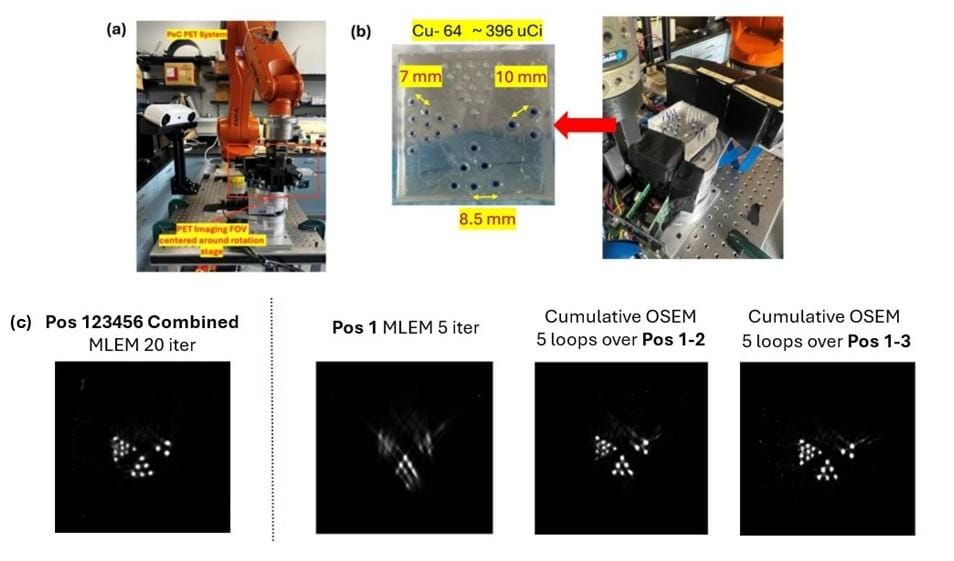
Using the portable point-of-care system, the study investigated the feasibility of interactive scanning and a real-time image updating strategy. Researchers imaged a phantom containing three clusters of radiotracer-filled rods while moving the detector panels to six user-selected positions.
Because data acquisition time was significantly longer than the reconstruction time, the system continuously updated images as data was acquired. This was compared to a conventional reconstruction framework where images are generated only after the entire scan is completed.
The study found that image quality from the real-time updating framework was comparable to the conventional framework. Phantom structures became clearly distinguishable after three to four positions, suggesting that clinicians could potentially terminate a scan early if the imaging tasks are fulfilled. Alternatively, image quality can be further improved with additional scanning positions or reconstruction iterations.
A Shift in Bedside Workflow
“This proposed approach better supports interactive and adaptive imaging workflows at the bedside,” says Xiyan Li, a graduate researcher in the imaging science doctoral program at Washington University in St Louis, in a release. “It represents a paradigm shift that offers new avenues to deploy novel molecular imaging applications.”
The current study utilized a benchtop prototype system for the point-of-care technology. Researchers are currently building a prototype system suitable for initial human imaging studies, which are scheduled to begin in 2027.
Photo caption: (a) Benchtop portable POC-PET prototype system. (b) Phantom imaging experiment setup. (c) Comparison of conventional full-data MLEM reconstruction with incremental OSEM reconstruction.
Photo credit: SNMMI, Abstract 262595. “Interactive PET Scanning and Real-Time Image Reconstruction for a Portable Point-of-Care PET System,” Xiyan Li, Samarth Aggarwal, Ling Cai, Pinhuang Wang, Richard Laforest, Joseph A. O’Sullivan, and Yuan-Chuan Tai, Washington University in St. Louis.