 |
· A Better Brain Picture
· Connect Imaging Customizes Its Mammo ViewBox
A Better Brain Picture
Wade Mueller, MD, says the biggest problem he and his peers in neuroscience face is that there are more streams of information about a patient compared to what was available in the past.
“The problem we have is organizing it all in one brain space,” said Mueller, a professor in the Department of Neurosurgery at the Medical College of Wisconsin.
A suite of three FDA-cleared brain scan software products from Prism Clinical Imaging helps Mueller overlay and intersperse different data sets from functional MRI, diffusion tensor imaging, and advanced physiological studies. The technology provides for image acquisition, postprocessing, clinical viewing, and export to treatment systems while interoperating with existing imaging modalities, PACS, DICOM infrastructure, and treatment systems in radiation oncology and neurosurgery.
“We can represent those images in a fashion that allows the clinician to vary the opacity or intensity of the images in relation to one another,” said Paul Schmelzer, Prism president and CEO. “Physicians could then see multiple characteristics of the patient’s condition in one single viewing space, which we refer to as a deck.”
 |
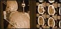
| Prism Clinical Imaging offers a suite of three FDA-approved brain scan software products for neurosurgeons and radiation oncologists. |
On the upper left corner of the viewer is a stack of bars—some gray, others white, and the remainder in color. Each of them corresponds to a volume of images that were acquired to highlight a particular characteristic. The Prism software allows the physician to overlay individual parts of data on top of one another, “almost like paging a book,” Mueller said.
“Once you can see the patient’s anatomy, location of pathology, and functional tissues that surround it, then you can devise a treatment plan that avoids functional structures and reduces the risk and incidence of side effects,” Schmelzer said.
Schmelzer explained that in a typical surgical planning situation, the neurosurgeon has both images side by side on a screen or lightbox, and they must envision the picture in their mind of how they will approach the tumor. “What we do is add additional information to the pictures,” Schmelzer said. “We let the computer put those two anatomical images together for the physician.”
Mueller cited a number of the software’s benefits, praising its user-friendly functions. “It’s very easy to use,” Mueller said. “You can stack multiple layers on top of each other, and you can extract one layer off just as easily. In the act of doing that manipulation, you can educate yourself better about this individual’s brain or brain problem.”
Schmelzer also mentioned another benefit of the software, comparing it to sole contrast agent-enhanced renderings of the tumor. In these renderings, physicians can view a binary, black or white, rendering of the boundary of the tumor. “But the reality of those tumor boundaries is that the metabolic signatures of the tissue may not correspond to those contrast agent boundaries,” Schmelzer continued. “In fact, there are early indications of the spread of pathology that a metabolic signature will pick up that a contrast agent will not. You get a better understanding of the tumor’s boundaries using physiological sequences than pure, contrast agent-based renderings.”
Moreover, the intuitive physician interface includes tools that are useful in validating data quality, identifying regions of interest, and highlighting overlapping activation patterns.
Formerly Kyron Clinical Imaging Inc, Prism Clinical Imaging was founded by practicing professionals at the Medical College of Wisconsin. As part of its marketing plan last year, the company recruited a medical advisory board and focused its installations in the academic arena. “We wanted to deploy our software in academic medical centers, where we can expose it to the thought leaders in the field and gain their appreciation for the value of the tool. More importantly, we can solicit their input to the further development of the system.”
—Elaine Sanchez
Tech Zoom
Connect Imaging Customizes Its Mammo ViewBox
Software Solution Tailored for Super-High-Res Display
Though Mammo ViewBox, the latest software module from Honolulu-based Connect Imaging, boasts additional capabilities to specifically support the technology of the Totoku MS51i2 21.3-inch digital mammography, super-high-resolution display, it is compatible with any 5MP display.
“The software, when used with the Totoku display, offers three times the horizontal resolution for viewing mammography images,” said Philip J. Manly, president and CEO of Connect Imaging. While the exact nature of the enhancements is confidential, he added that major changes were required to the display portion of the workstation software to support the Totoku super-high-resolution display. “This is most important for viewing screening studies, because the radiologist does not know what, if anything, he will find.”
The solution is at its best scanning the initial display of such studies (4×2, eight images on two monitors including the prior study), when the mismatch between image resolution and screen resolution is greatest.
“Increasing the horizontal resolution results in better definition of microcalcification—sharper edges and more accurate shape—[and] because there is less averaging of microcalcifications with surrounding tissue, the contrast between them is greater,” he said.
The Mammo Viewbox workstation is a full diagnostic workstation software, automatically hanging studies without user intervention. The specialized solution allows clinicians to determine how images are viewed and to manipulate images without looking away from the display. A number of quick-access tools also have been built into the system, including the ability to immediately put the study in diagnostic reading mode with a double-click of the mouse.
Customizing a version for Totoku is an example of Connect’s ability to respond quickly to customer requests, according to Manly. “Because many of our clients utilize complex workflow, Connect Imaging has developed the capacity to handle customization quickly. Within a few weeks of receiving engineering samples of the Totoku monitor, we had implemented the changes,” he said.
US Electronics Inc, Totoku’s leading worldwide distributor, was appreciative of the effort. “We believe this display is a significant improvement over standard 5MP mammography displays, even those that can display 10 bits of grayscale,” said Madhu Reddy, president of US Electronics. The MS51i2 display is equipped with independent subpixels drive (ISD) technology, allowing it to accept higher-resolution output from modality devices. “We talked with several PACS companies about the software changes that would be necessary to support the higher resolution. Connect Imaging responded immediately, and is now the first PACS company to release software that supports our new monitor.”
Those without the Totoku display are still able to take advantage of the improved software. “We now can offer this workstation software to all users of digital mammography,” Manly said. “Though it works most efficiently when combined with other mammography modules in the Connect Imaging PACS, our Mammo ViewBox workstation software is compatible with any DICOM archive.”
The Mammo ViewBox software currently supports the major elements of the Integrating the Healthcare Enterprise mammography profile, and will support all the elements before the end of summer, according to the manufacturer.
In addition to the Mammo ViewBox, Connect Imaging offers four separate products designed to optimize the workflow in the mammography practice. The company’s mammographer workstation provides mammography technologists with easy access to priors and to the radiologist’s annotations so they can plan the views they take.
The entire product line is designed to support flexible image flow that can be adapted to a facility’s preferred way of handling the more complex workflow of diagnostic studies, including incremental views, according to Manly.
Connect Imaging also offers a version of its ViewBox technology suited to handle other, nonmammo modalities. Among other features, the ViewBox makes switching between image mode and series mode easy work; series, including those from prior studies, also can be linked for simultaneous scrolling.
Clinicians can define a different hanging protocol for each modality, and all configurations are maintained separately by the user. The ability to perform 3D reconstruction functions also is available with the ViewBox, including MPR, MIP, MinIP, and volumetric reconstruction using predefined templates.
—Dana Hinesly
