Researchers have developed an AI tool that mimics radiologists’ gaze patterns using heat maps to improve chest X-ray analysis.
Artificial intelligence (AI) can scan a chest X-ray and diagnose if an abnormality is fluid in the lungs, an enlarged heart, or cancer. But being right is not enough, says Ngan Le, PhD, a University of Arkansas assistant professor of computer science and computer engineering. We should understand how the computer makes its diagnosis, yet most AI systems are black boxes whose “thought process” even their creators cannot explain.
“When people understand the reasoning process and limitations behind AI decisions, they are more likely to trust and embrace the technology,” Le says in a release.
Le and her colleagues developed a transparent, and highly accurate, AI framework for reading chest X-rays called ItpCtrl-AI, which stands for interpretable and controllable artificial intelligence. The team explained their approach in a study published in the current issue of Artificial Intelligence in Medicine.
Incorporating Gaze Heat Maps
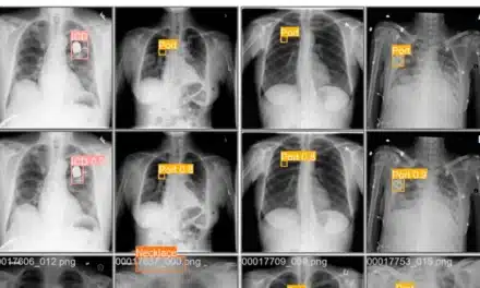
The researchers taught the computer to look at chest X-rays like a radiologist. The gaze of radiologists, both where they looked and how long they focused on a specific area, was recorded as they reviewed chest X-rays. The heat map created from that eye-gaze dataset showed the computer where to search for abnormalities and what section of the image required less attention.
Creating an AI framework that uses a clear, transparent method to reach conclusions—in this case a gaze heat map—helps researchers adjust and correct the computer so it can provide more accurate results. In a medical context, transparency also bolsters the trust of doctors and patients in an AI-generated diagnosis.
“If an AI medical assistant system diagnoses a condition, doctors need to understand why it made that decision to ensure it is reliable and aligns with medical expertise,” Le says in a release.
Need for Transparent AI Framework
A transparent AI framework is also more accountable, a legal and ethical concern in areas with high stakes, such as medicine, self-driving vehicles, or financial markets. Because doctors know how ItpCtrl-AI works, they can take responsibility for its diagnosis.
“If we don’t know how a system is making decisions, it’s challenging to ensure it is fair, unbiased, or aligned with societal values,” Le says in a release.
Le and her team, in collaboration with the MD Anderson Cancer Center in Houston, are now working to refine ItpCtrl-AI so it can read more complex, three-dimensional CT scans.
Photo caption: Ngan Le, assistant professor of computer science and computer engineering, studies AI and computer vision.
Photo credit: Russell Cothren
Summary:
Researchers have developed ItpCtrl-AI, an artificial intelligence (AI) tool designed to analyze chest X-rays using gaze heat maps that track how radiologists examine images. By incorporating this data, the AI system prioritizes key areas for identifying abnormalities such as fluid in the lungs, an enlarged heart, or cancer. The approach aims to improve diagnostic accuracy while making the AI’s decision-making process more transparent. Researchers are now working with MD Anderson Cancer Center to adapt the system for analyzing 3D CT scans.
Key Takeaways:
- AI Model Uses Radiologist Gaze Data – ItpCtrl-AI was trained on eye-tracking data from radiologists to determine where to focus when analyzing chest X-rays.
- System Designed for Transparency – The AI framework provides a visible reasoning process, allowing clinicians to assess how it arrives at diagnoses.
- Researchers Expanding Capabilities – The team is working to refine the system for use with 3D CT scans in collaboration with MD Anderson Cancer Center.